Analytical techniques have come a long way, but what does the future hold? Rachel Brazil asks the experts what they’d like to see
-
Resolution and sensitivity: Current analytical techniques like spectroscopy and crystallography have limitations in resolution and sensitivity. Advances such as electron diffraction and improved detector technology are helping to overcome these challenges, allowing for the analysis of smaller crystals and more precise measurements.
-
Differentiating complex molecules: Techniques like ion mobility spectroscopy are being developed to better differentiate between similar molecules, such as isomers and chiral molecules. Innovations in this area could significantly improve the analysis of complex organic molecules and polymers.
-
Real-time analysis: There is a growing need for real-time analysis of chemical reactions and materials. Techniques like hyperpolarisation in NMR and advances in x-ray spectroscopy are being explored to monitor reactions as they happen, providing insights into reaction mechanisms and material changes.
-
In situ analysis: Analysing molecules and materials in their natural environments remains a significant challenge. Techniques like cryo-electron microscopy and computational methods are being used to study biological molecules in more realistic conditions, but further advances are needed to achieve accurate in vivo analysis.
This summary was generated by AI and checked by a human editor
Chemistry is often seen as synthesising new molecules and materials, but being able to analyse what you have made is just as important. The wet chemical tests and titrations of yesteryear have now largely been replaced by spectroscopic or crystallographic methods, probing elemental composition, crystal structure, molecular mass or nuclear spin. We’ve come a long way since Robert Bunsen and Gustav Kirchhoff’s first flame emissive spectroscope in 1860, which they used to identify elements, even discovering a few along the way (rubidium, caesium, thallium and indium – all named after the colours observed). We now have methods that can detect parts per billion quantities and can image some molecules at the angstrom scale.
But have we come far enough? Is our ability to analyse keeping up with the progress synthetic chemists are making? Can we probe mixtures or analyse reactions progressing in real time or follow functional materials changing as they perform? And what about larger biological molecules, whose structures need to be understood in their natural environments. Today’s chemists still have a wish list of things they would like to analyse, but are beyond our current capabilities.
Seeing smaller

At some point resolution and sensitivity are limitations for almost all analytical techniques. Lucy Saunders, beamline scientist at Diamond Light Source in Didcot, UK, says researchers are often seeking to detect very minute contaminants in their samples with the various spectroscopic methods they can employ at Diamond. ‘On our beamline, we have some very high-resolution detectors, and they have been used to detect a limit of about 0.01% of a component.’ Julia Parker, her colleague at Diamond, says sensitivity varies depending on the technique. ‘You might be able to look at things down to parts per million concentrations.’ She says that, when looking at imaging, ‘we are mapping with a 50nm beam, and that’s the limit of the spatial resolution that you can currently achieve using these techniques.’ This isn’t always good enough though. ‘If you’re looking at how molecules are interacting with a larger matrix, or you’re looking for active sites within a catalyst material, you want to really be able to look at them on smaller length scales,’ says Parker.
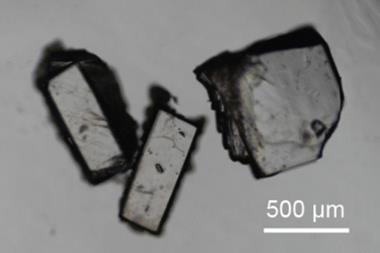
There is also a scale problem with x-ray diffraction, the technique used to probe crystal structure. ‘No matter how powerful you make your x-rays, the technique can only work on a finite sized crystal,’ says Simon Coles, who is a structural chemist at the University of Southampton in the UK and director of the UK National Crystallography Service. The lowest end of the scale for crystal size is 1–5µm. ‘Quite a lot of time and effort has been put in over a number of decades to try and get round that and look at smaller particle systems. Powder diffraction does that, but it’s very difficult to get the same resolution answer – it’s almost impossible, says Coles.
In the last 5–10 years, there has been a breakthrough – the answer has been diffracting electrons rather than x-rays, using a modified electron microscope. ‘Electron microscopes have been around for donkey’s years, and have been able to do diffraction, but its only now that we have good detector technology,’ says Coles. Current semiconductor hybrid pixel detectors can count every photon as it arrives, making them quicker and more accurate, providing structural information from crystals in the tens and hundreds of nanometer range. ‘That opens up the technique to whole areas of chemistry,’ says Coles.
But it hasn’t yet solved all problems. Electrons interact very strongly with matter and produce secondary, tertiary and even quaternary diffraction effects, making the data sometimes difficult to interpret. ‘There’s still quite a lot of method development to go,’ says Coles.
Spot the difference
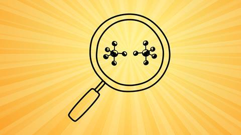
Being able to better differentiate similar polymers and large organic molecules would be on the wish list of mass spectrometry specialist Perdita Barran from the University of Manchester in the UK . ‘A lot of the commercially available synthetic polymers are difficult to analyse with most mass spectrometry methods,’ she says. They can have very similar structures, with perhaps only one CH2 unit difference and their size makes them difficult to separate chromatographically.
Barran says there can also be issues with molecules such as glycans and lipids where different structures can be isobaric species (identical mass) or isometric species (same formula, different structure). Things become very complicated in molecules with regio or stereo isomers, where mass spec really cannot differentiate at all.
One recent innovation that might change this is a new way of using ion mobility spectroscopy. ‘Whenever your laptop’s swabbed in an airport, it’s being taken to an ion mobility spectrometer,‘ says Barran. It measures the movement of an ion through a gas-filled tube and the arrival at the detector then is based on how mobile that ion is in the gas. Shape becomes a differentiating factor as well as mass and charge which allows the separation of some structurally different isomers.
But the trickiest problem is still chiral molecules. ‘Enantiomers are the thing that we really need to sort’ says Barran. In February, chemist Zheng Ouyang from Xinhua University in China proposed a method that might solve this problem. By adding a weak electric field to the ions as they travel through the gas, the molecules start to spin and this gives each enantiomer a slightly different collision path. Barran says this new method is ‘really promising’ – but it’s early days.
Real-time analysis

Another issue is analysing complex mixtures or composite materials. It’s a problem for almost all current techniques. For example, Barran says things like germanium or antimony nano dots can be hard to analyse. ‘These are super massive things that are often polymerically coated. So whilst they are perceived as being quite homogenic with respect to their functional properties, they’re actually quite disparate.’
NMR chemist Jean-Nicolas Dumez is also looking for better way to analyse mixtures, to speed up identification and cut out lengthy purification processes but also to analyse species that cannot be isolated from a reaction. ‘So if one wants to characterise them, they need to characterise in the reaction mixture,’ he says. Dumez, from the University of Nantes in France, is developing more sensitive NMR methods to be able to interpret signals from multiple species in one scan. It is already possible to do this, provided that they have well-resolved and previously identified peaks in the spectrum, but peak overlap can get in the way and prevent quantification of different compounds. ‘My personal dream would be being able to have the data that is equivalent to what you would have for pure compounds, but directly from the reaction mixture and for as many compounds as possible,’ says Dumez
The real prize would be to do this in a dynamic way – to be able to analyse chemical reactions in real time, to better understand reaction mechanisms or to follow crystal growth. Parker says that at Diamond these types of capabilities depend on how fast you can scan, the efficiency of your detectors and the intensity of the x-ray beam. She is trying to probe biomineralisation processes and one of the problems is the interference from the liquid solvent which is inevitably present.
Monitoring reactions as they progress with NMR currently also has limitations. ‘One has to be able to collect data on a time scale that is relevant for the reaction of interest, and [with high] sensitivity, because sometimes species of interest will have low concentration,’ says Dumez. At present real time NMR cannot follow reactions faster than a few seconds with a limit at around 50ms time resolution.
Some new methods are improving NMR sensitivity, one being hyperpolarisation, which means samples are prepared so that the spin states are not at thermal equilibrium and more of the molecules present will be able to contribute to the signal. This can be done in several ways, one being para hydrogen induced polarisation, which chemically incorporates polarised hydrogen atoms into molecules. Another is dynamic nuclear polarisation, where the sample is cooled to extremely low temperatures in a strong magnetic field and exposed to microwave radiation. ‘But it’s not yet the case that you can polarise anything, or that you can polarise it for long enough, so clearly, there’s still progress to be made,’ says Dumez.
Our technique historically has required the crystal to be very still – it’s like Victorian photography, you couldn’t capture movement
The sort of spectroscopic techniques available at Diamond can also provide real-time analysis, but there are limitations too. Parker and Saunders are hoping that the ongoing Diamond 2 upgrade will improve their capabilities for real-time analysis. The new fourth generation light source technology will provide much brighter photon beams and an increased coherence, meaning the photons are more in phase. ‘That will then help with focusing down to smaller beams to improve that spatial resolution and also to do things much faster, so you’re able to look then at more short-lived reaction intermediates,’ explains Parker. Diamond 2 will allow them to push down to 10nm resolution and will also enable new imaging techniques like ptychography, a type of coherent diffractive imaging which uses a coherent x-ray beam to retrieve the phase information from diffraction patterns, producing better spatial resolution than conventional optics.
The Diamond beamline will shut down from late 2027 for 18 months for the upgrade, but one proposed beamline on their wish list called Berries (bright environment for x-ray Raman, resonance inelastic and emission spectroscopies) won’t be going ahead due to financial constraints. Many chemists are still lobbying for it. The beam line would have provided pink-beam x-ray emission spectroscopy (pink-XES; a pink x-ray beam is focused using mirrors to more narrowly filter the energy). Pink-XES is an element-selective technique covering transition metals, a number of other metals and light elements, and can provide information on the environment of an atom, for example the ligands a metal is bound to, or an atom’s oxidation state. It’s ideal for studying catalysts and the changes occurring over the course of their use and it’s able to collect data quickly, opening the door for time-resolved studies. But it’s something UK chemists will have to wait a little longer to access.
Crystallographers are also looking for dynamic methods, particularly to study the sorts of porous materials now being developed for hydrogen or greenhouse gas storage, or solid-state materials with electronic properties such as OLEDs used in TVs. ‘Our technique historically has required the crystal to be very still… it’s [like] Victorian photography, you couldn’t capture movement,’ says Coles, but ‘what we [now] need is video.’ On Coles’s wish list would be a technique with the precise geometry of atomic structures you get from crystallography, but with the ability to track changing structures.
He says there are the beginnings of such methods with the x-ray free-electron laser (XFEL) which generates 100fs pulses of intense, coherent x-ray beams which provides multiple datasets showing changes in chemical bonding or vibrational energy that can be pieced together like a film. It’s an ideal technique to look at biological samples such as membrane proteins, and the technique has solved the atomically resolved structures of several important G-protein coupled receptor proteins. There are very few of the instruments globally and Coles says it’s very early days with dynamic measurements which are still largely the preserve of spectroscopy. ‘That’ll be another technological step change in decades to come, I would expect,’ he says.
In the real world

The other big challenge for chemical analysis is being able to observe molecules and materials in situ – in the environments they are being used. ‘It’s a massive challenge that we really can’t do,’ says Coles. One of the most difficult environments to replicate is biology. Biophysicist Mahmoud Moradi from the University of Arkansas in the US has been looking at this challenge for protein structures. ‘We do know that proteins are very sensitive to the environment that they live in,’ he says. But in most analytical methods you have to take the protein out of its normal physiological environment and put it in a controlled condition, usually based on the technique being used.
Moradi is particularly interested in cross-membrane proteins and for x-ray crystallography these proteins are often placed in a detergent solution to try and replicate the cell phospholipid membrane. ‘Something that people have realised more recently is it makes a very big difference [to the structure],’ he says.
You may have to work on a protein for years to be able to eventually crystallise it
Current techniques often leave parts of these protein structures unresolved, such as cytoplasmic loops which are functionally and structurally important for drug design. Data on these types of feature and how they interact with a natural membrane are often just missing. Some researchers have used artificial phospholipid membranes which are more representative. ‘But even if you do that, it’s not going to be the physiological lipid environment of the protein,’ explains Moradi. ‘So that’s the biggest challenge… I don’t really know of any structure determination technique that works in vivo!’
The difficulty of crystallising proteins for crystallography is a very old problem and still requires a lot of tricks, including adding mutations or even parts of other proteins. ‘You may have to work on a protein for years to be able to eventually crystallise it,’ says Moradi. But the advent of cryo-electron microscopy (cryo-EM) has provided another route. ‘You don’t have to put [the protein] in a particular condition that crystallises it.’
The technique involves flash-freezing solutions of proteins or other biomolecules and imaging them with an electron microscope, which can achieve a resolution of about 3Å. But cryo-EM still has a lot of limitations. ‘In terms of the environment, it has the same flaws: you still [need to] do this in vitro, so you still choose the same type of environments, [and] it’s cryogenic, so you need to go to very low temperatures and that itself is a problem,’ says Moradi.
He is now concentrating on computational methods for simulating proteins in physiological conditions, starting with data collected from crystallography or cryo-EM. The hope is the model will correct any changed introduced. ‘If we’re lucky enough, and we can run long enough simulations, we may be able to find the stable conformation of the protein and its dynamics in physiological conditions, but just like crystallography the computational approaches also have their own limitations,’ acknowledges Moradi, as they can’t yet accurately model the complexity of a biological cell membrane.
For those probing biology the next frontier is analysing the chemistry of the single cell and understanding its chemical make-up quantitatively, from small molecule metabolites to large proteins and other biomolecules. There are already attempts to do this, including single cell mass spectrometry, which uses an electrospray ionisation technique. Rather than fragmenting large proteins, it turns them into smaller and smaller droplets until they become molecular ions which will travel through the mass spectrometer. It’s not new but Barran says we are now getting to a point where we can differentiate between different cell types, look at how cell protein levels change when a drug is introduced or the difference between cancer or healthy cell. ‘I’ve been very impressed by the evidence at an early stage,’ she says.
Our ability to analyse molecules and materials has played a huge role in the advances chemists have made, but there are still things we cannot detect, measure or image. As new techniques are developed, we are likely to burst through the current boundaries which will be replaced with new ambitions for analysing on a smaller scale, at faster rate, and in more authentic and challenging environments.
Rachel Brazil is a science writer based in London, UK








No comments yet