Biomaterials engineers are hijacking the chemistry behind the healing process to create a new generation of smarter wound treatments. Victoria Gill investigates
Biomaterials engineers are hijacking the chemistry behind the healing process to create a new generation of smarter wound treatments. Victoria Gill investigates
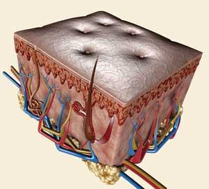
There’s one human organ more at risk of damage than any other - constantly exposed to the dangers of everyday life. It cushions, protects and insulates your body against the elements, and against infection. And it’s constantly active: regulating temperature, synthesising essential vitamins and pigments, regenerating and healing. It’s your skin.
And damage to the skin - so easily inflicted - can be devastating. Injuries that are difficult to heal can leave disfiguring scars, and worse, open wounds provide an ideal site for infection to take hold - particularly dangerous in an era when the prevalence of multi-drug resistant bacteria is on the rise.
The next generation of wound-healing technologies will work with the body’s own ability to regenerate. Through a combination of chemistry, materials design and a thorough understanding of the biology of healing, new biomaterials that improve the healing process itself are gradually moving from the laboratory towards the clinic.
The aim is to design and build something safer, cleaner and more efficient than nature can manage.
’We still have more to learn to fully appreciate the complexity of biology,’ says Achim G?pferich, head of a research unit in the department of pharmaceutical technology at the University of Regensburg, Germany. ’But we have reached a stage now where we can start to tailor materials to the needs of the body.’
Tissue engineers and biomaterials developers have spent decades looking for ways to get new cells to a site of injury. According to G?pferich, an over-simplified approach has been the downfall of many projects.
’I think we underestimated the impact of biology on tissue engineering. Many people thought: "we have materials that cells will grow very well in so we can use those to deliver the cells and they will integrate into the tissue". There was some success with that approach, but there were also many failures.’
Now researchers are focusing on more than cell delivery and new materials will deliver a whole host of healing agents, including growth factors and antimicrobials, directly to an injury. ’You need to deliver the right agent at the right time,’ says G?pferich. ’If you have a material that delivers something very effectively, but it misses the stage during the healing process when that agent is most needed, that’s just a waste.’
Refine and replace
’In some cases, we started using biomaterials simply to provide an alternative for natural materials,’ says Sheila MacNeil, professor of tissue engineering at the University of Sheffield in the UK. ’But, as you go down that route, you realise you can fine-tune a material to give you advantages and give a lot more control over wound healing.’
Human skin is composed of three layers. The uppermost epidermis contains a tightly integrated sheet of barrier cells called keratinocytes. This is attached to the dermis, which is made up mostly of collagen - a strong, fibrous protein - and contains blood vessels and sensory receptors, as well as sweat glands and hair follicles. The subcutaneous layer is connected to the dermis by elastin and collagen fibres, and is made up largely of adipocytes - cells that accumulate and store fat.
In the 1980s, clinicians began to treat burns patients using sheets of keratinocytes cultured from the patients’ own skin to replace the damaged epidermis. Where the patient’s dermal collagen is destroyed it is often replaced using animal-derived materials. Cadaver skin from tissue banks can be used but this is in limited supply. Extracted porcine (pig) collagen, and, more commonly, bovine collagen are often used in cases where the dermis is very badly damaged.
But there has long been a move to develop safer substitutes not taken from animal sources, particularly in the wake of the BSE crisis in the UK. ’With human cadaveric tissues you can screen them for viral and bacterial disease and you can get them to the same safety level as a blood transfusion, for example. But with BSE, there’s no simple screening test so there’s a drive to develop safer alternatives that aren’t animal-derived,’ says MacNeil.
So a number of research teams, including MacNeil’s own, started working with polymers, with the aim of developing safer, more convenient alternatives.
MacNeil’s team started with polylactic acid (PLA) and polyglycolic acid (PGA) - already approved for resorbable surgical stitches.
’There are lots of brilliant polymer chemists who can create all kinds of different materials with different properties, so there’s almost no limit to what you can dream up,’ says MacNeil. ’But some people say there’s no need for any more new polymers for these applications because the bottleneck happens in the regulatory process. If you’re serious about getting into the clinic, you’ll have a much better time if you start off with a material that’s already approved.’
MacNeil’s team builds polymer scaffolds that can be seeded with cells or molecules that promote healing - integrating a structure that supports the wound with direct, topical treatment. This is done by electrospinning the polymers - passing the viscous polymer liquid through a conducting needle to form a thread. The difference in voltage between the charged conducting needle and an earthed collector below it creates an electric field that draws the liquid through the needle. It’s a very useful processing method for biopolymers.
In an ongoing collaboration, MacNeil and her colleagues at the University of Sheffield - polymer chemists Steve Rimmer and Linda Swanson, and microbiologist Ian Douglas - are developing a new polymer which will light up when a bacterial infection is present in an open wound.
This polymer is based on poly-(N -isopropylacrylamide) or PNIPAM, which Rimmer describes as ’probably the most studied, thermally responsive polymer in the world’.
’We use reversible addition-fragmentation chain transfer [or Raft] polymerisation to make a highly branched polymer,’ he explains. ’When you do this you get lots of end chains and we can modify these with ligands for whatever we want to attach, in this case antibiotics.’
By coupling the branched polymer to two antibiotics - vancomycin and polymixin-B - the team created a material that bacteria readily bind to. It can identify the two major classes of bacteria: vancomycin binds gram positive bacteria and PMX-B binds gram negative bacteria. (These two classes have structural differences in their cell walls and require treatment with different classes of antibiotic, so it’s clinically important to distinguish them.)
In lab studies, increasing the temperature above a critical point causes the thermoresponsive material to collapse into an insoluble globule, principally because hydrogen bonds within the polymer and between the polymer and the water are broken. ’This is the clever bit,’ says MacNeil. ’If the bacteria are on the end of the chain, you can drag them out of solution too.’
The team was able to change the temperature at which the polymer collapses to suit the application. ’If we produce highly branched polymers, we can control the transition temperature,’ says Rimmer. ’The structure of the end group is important in determining this temperature. The binding event involves a charged ligand locking into a binding pocket, which perturbs these end groups.’
So the team was able to make a polymer that changes transition temperature when the bacteria bind. And by designing the polymer so that this new transition temperature is lower than normal body temperature, the team can ensure that the binding itself causes the polymer to collapse and drag the bound bacteria out of solution.
’With a branched material we can also use luminescence to see what’s happening,’ says Rimmer. Using two fluorescent labels - which are added to the mix before the monomer is polymerised - his team has been able to monitor the structural changes. ’There are two labels on the polymer and they talk to each other,’ he says. (The group’s exact method for attaching the fluorescent polymer is under wraps pending a patent.)
’The fluorescent labels work via non-radiative energy transfer,’ explains Swanson, a specialist in photophysics. ’You have a donor and an acceptor and they have to be within 10nm of each other to generate a signal. So in the expanded conformation, where the chain ends are more than 10nm apart, there’s no signal, but in the collapsed conformation there is.’
’Part of our funding comes from the UK defence department,’ says MacNeil. ’This could be helpful in a military field hospital - with a fluorescent label bound to the polymer, you could just shine a fluorescent lamp on a wound and detect if it’s infected. ’My son is in the Marines, so my interest in this is quite personal,’ she adds.
The team has also managed to harness a key healing process within a new hydrogel material, which binds to heparin, a compound that is released by blood vessels at a site of injury. Heparin mediates the release of growth factors in the body, which promote tissue healing.
’Many growth factors don’t work without heparin,’ says Rimmer. ’We’ve developed a hydrogel that contains a peptide modelled on the binding region of vascular endothelial growth factor. So that site will bind heparin.When this is tested in the lab the hydrogel binds heparin and the heparin binds a growth factor which causes new blood vessel formation.’
The material is being developed so that it will bind the patients’s own heparin and growth factors in the body, keeping them where they are needed to speed up wound repair.
Polymers of life
Suwan Jayasinghe, a biophysicist at the UK’s University College London, has combined tissue engineering, regenerative medicine, and polymer processing in a single step. Jayasinghe’s team was the first to seed polymers with living cells using the electrospinning method. Mainly working with polyethylene glycol and polymethyl methacrylate systems, he adds a medium containing a wide range of cells - from immortalised human tissue cells to embryonic stem cell cultures - before the polymer is drawn through a needle to form a thread that can form a living scaffold. This technique is called ’cell electrospinning’.
’We’ve found out already that once the cells have undergone electrospinning, they are undamaged,’ says Jayasinghe. He suggests that the technique could eventually produce a living scaffold tailored to every individual patient. ’That’s a huge challenge: we might need to develop a different biopolymer for each different cell type. Our next step is to find out how these scaffolds behave in an animal model,’ he says.
His team has created another new processing method - aerodynamically assisted jetting - to form foams and emulsions that contain living cells.
They mixed alginate - a viscous copolymer extracted from seaweed - with phosphate-buffered saline or cell media containing embryonic stem cells. The liquid then flowed into a chamber while gas was passed through a needle inside the same chamber. Jayasinghe calls the technique ’microbubbling and microfoaming’. ’The flow of media into the chamber causes a differential pressure or flow field which draws out the gas flowing within the needle,’ he explains. ’If both the chamber and the needle were holding liquids then we could generate mixed liquid droplets to encapsulate cells and/or drugs.’
Back to nature
Ron Raines, a biochemist from the University of Wisconsin, US, has taken a novel approach to the problem of wound healing by copying nature but adding a chemical twist. His team has been working with a form of synthetic collagen which he first successfully synthesised in 2006 (see Chemistry World , April 2007, p43). Since then Raines has been using animal pelts to investigate the collagen’s use in wound healing.
In the skin, collagen fibres form triple helices, but Raines’ synthetic form is subtly different. He ’decorates’ single collagen strands with bumps along their surfaces (the details of how these bumps are engineered are still under wraps). ’It’s all about steric effects,’ says Raines. ’These strands won’t form triple helices with one another, but they will with the collagen in the wound.
’In fact, the damaged collagen seems to love them - it forms very strong bonds and triple helices. We take advantage of the fact that in a wound, collagen is trashed - there are lots of binding sites where it’s been damaged and our strands preferentially bind at those sites.’
So far his team has attached dye molecules to the cell strands to show up the binding. And he suggests that this investigative method has diagnostic potential.
’Imagine putting a collagen-bound fluorescent dye in a spray bottle. A surgeon could spray it onto the wound to see where the collagen is damaged the most. This would show them where they need to pay the most attention and also show up the area most at risk of developing scar tissue,’ says Raines.
The next step is to attach other molecules that promote healing to the collagen strands - growth factors, peptides or other small molecules. ’We’re very excited about this. Again it’s taking advantage of the dynamics to recruit these agents to the place they’re needed most.’
Raines says that by understanding the chemistry of such important biological molecules, he and his team are able to take a ’more atomic approach’ to treating wounds. And, like MacNeil, he has also considered the pathway to the clinic in his laboratory work. ’Our material is very biomimetic. We’ve not tested the immunogenicity yet but it’s so similar to natural collagen that it’s unlikely to cause problems. And topical treatments tend to have a much easier time, so the road to the clinic shouldn’t be too bumpy.’
Next generation
One US company - Florida-based Quick-Med - has already thrown down the regulatory gauntlet. As Chemistry World went to press, the company was expecting imminent FDA (US Food and Drug Administration) approval of its new antimicrobial wound dressing, known as Nimbus, which stands for Novel Intrinsically MicroBonded Utility Substrate.
The basis of the dressing is a large polyquaternary ammonium compound - poly(diallyl dimethyl ammonium chloride) or poly(DADMAC). Because of their high positive charge, the numerous quaternary amine groups on the polymer inactivate or kill bacterial cells by disrupting the cell membrane and cell wall.
The polymer is irreversibly bound to a gauze dressing. ’The size of the polyquats [we use] is such that it continues to kill in the presence of body fluids, such as blood, serum, urine and perspiration,’ explains Jerry Olderman, vice-president of R&D at Quick-Med.
Gregory Schultz, a clinical researcher from the Institute for wound research at the University of Florida, US, was one of the team that developed the polymer coating, and licensed the intellectual property to Quick-Med. He is particularly excited about what the approval of Nimbus means for the future of wound-healing technologies.
’Traditional wound dressings absorb fluid from the wound and act as a great incubator for bacteria that are shed back into the wound,’ says Schultz. ’This is the first bound antimicrobial material - rather than delivering an antimicrobial into the wound, it creates a barrier that won’t allow bacteria to penetrate the dressing or to grow in the wound fluid.’
The FDA is treating Nimbus as a new medical device, which means that it will be considered to be without precedent and will receive de novo approval rather than going through the standard 510(K) clearance, which is used for medical devices that are essentially an improved equivalent to something already on the market. There have been only 36 de novo approvals of new medical devices in the past 11 years, compared with about 3000 approvals per year for 510(K) clearance, says Schultz. ’It’s a longer and more expensive approval process, but having survived that, the company can use this as a predicate for new dressings - attaching drugs to the same polymer coating for example.’
Quick-Med’s scientists are already developing the same dressing-bound polymer for the sustained release of molecules. ’A portion of the positively charged [amine] groups can be used to temporarily hold and release anionic [negatively charged] materials,’ says Olderman. ’In the late stages of development at the moment we have the polymer containing a protease inhibitor.’ This inhibitor blocks the action of the protease enzymes, which are highly elevated in chronic wounds and destroy collagen and other proteins that are essential for healing, slowing the healing process.
Schultz still works closely with the Quick-Med team and has received funding from the US military, which is interested in developing the technology for use in the field. ’The military is a good source of short term funding. They’re always extremely focused on exactly what they want,’ says Schultz.
’The success stories in this field always come from really integrated teams,’ says Schultz.
As biology and chemistry are cleverly combined to understand a wound’s molecular environment, biomaterials are designed to manipulate it. Perfect teamwork.
Victoria Gill is a science journalist based in Lancashire, UK
Further Reading
S Carter, B Hunt and S Rimmer, Macromolecules , 2005, 38 , 4595
S Rimmer et al, Soft Matter , 2007, 3 , 971


No comments yet