Raman spectroscopy is no longer an insensitive technique. Tom Westgate finds out how this advancing technology offers new possibilities in biology and security
Raman spectroscopy is no longer an insensitive technique. Tom Westgate finds out how this advancing technology offers new possibilities in biology and security
’You need an information-rich technology to explore the nanoworld,’ says Renato Zenobi of the ETH Zurich, the Swiss Federal Institute of Technology. You might be forgiven for not immediately thinking of Raman spectroscopy as an example of such a technology, but Zenobi and many other researchers around the world would disagree. ’There are existing tools available, such as electron microscopy, atomic force microscopy and scanning tunnelling microscopy, but they give you no chemical information - a microscopy image can show you beautiful structures, but you don’t know what they are,’ he says.
Zenobi’s team is developing new techniques to increase the sensitivity of Raman. He suggests that of all the techniques that provide chemical information, Raman is the one that also offers high spatial resolution.
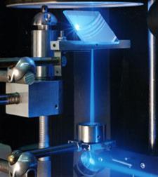
And in recent decades, the development of lasers and CCD (charge coupled device) detectors has produced step changes and brought smaller, faster instruments. Modified Raman techniques have already reached the fundamental limit of chemical detection - single molecules. Now, its ability to identify single biomolecules and cells could see Raman taking a frontline role in the molecular biology lab or hospital, and in airport security.
Drug companies have taken an active interest in developing Raman as an anti-counterfeiting measure. Scientists from Pfizer and the Science and Technology Facilities Council (STFC) Rutherford Appleton Laboratory in Oxfordshire, UK, have collaborated to develop a Raman-based method that can quantify the active pharmaceutical ingredient within intact drug capsules.
The technique is based on spatially offset Raman spectroscopy (SORS). An offset detector picks up photons that have travelled through the drug. Last year scientists reported that the technique could be used to analyse drugs within their packaging (see Chemistry World, March 2007, p9) and STFC scientists are investigating its use in a wider range of applications, including the detection of explosives in containers and the non-invasive diagnosis of bone disease and cancer.
Coming to the surface
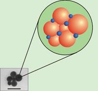
For Raman spectroscopy to reach into the realm of single molecule detection required the accidental discovery, in 1974, that molecules of pyridine on a rough silver surface gave an unexpectedly strong Raman scattering signal.
The fortunate discoverer of so-called surface-enhanced Raman spectroscopy (SERS) was Martin Fleischmann, who would later become embroiled in the cold fusion controversy. Fleischmann and his collaborators confirmed the signals were enhanced by a factor of a million. Raman had, until then, been burdened by its insensitivity to very low levels of a sample. Three decades later enhancements as high as 14 orders of magnitude have been reported, allowing the spectrum of a single molecule to be collected.
For the SERS effect to be observed, the molecule must be adsorbed to the surface of a colloidal nanostructure, generally of gold or silver atoms. When the laser hits the metal substrate it creates an enhanced local optical field. The molecule therefore feels an enhanced excitation which boosts the intensity of the Raman signal.
Katrin Kneipp of Harvard University and Massachusetts Institute of Technology, in the US, has been investigating SERS for over 20 years. She believes SERS has changed the perception of Raman. ’It was never used as a tool for trace detection because the Raman effect is too weak’ says Kneipp. ’Now, since SERS, people look at Raman in a new way - as a sensitive tool.’
Detecting single molecules holds fundamental interest for chemists and physicists, and to be able to identify them from a Raman spectrum makes SERS a promising technology for security applications, or, as Kneipp puts it: ’whenever you want not only to detect a single molecule, but also find out: what molecule?’
As well as picking up spectra from single molecules, Kneipp is applying SERS to biochemical analysis. Her team has introduced gold nanoclusters, measuring about 50 nm, into live cells. They have been able to record the SERS signatures arising from molecules on the surfaces of the particles in different compartments of the cell. ’The particles can be considered as tiny nanosensors’ said Kneipp. ’We can use them to probe the chemistry in the live cell.’
The rich variety of chemistry in different cellular compartments produces spectra that are difficult to interpret. To make sense of the complex data, Kneipp’s group compares the differences between spectra obtained as the nanoparticles move around the cell. As well as leading to an understanding of the cellular chemistry, Kneipp and others believe that the technique has potential for use in nanoscale biosensors that could be implanted and used to measure, for example, diabetes patients’ glucose levels.
Before this promise can become reality, however, SERS researchers must address the question of reproducibility in their measurements. The enhancement effect is strongest at a few ’hot spots’ on the surface, which have particular nanoscale features. But the number and location of hot spots is difficult to predict when making a new substrate.
The Raman Effect
The Raman effect was first reported 80 years ago in a paper in Nature on 31 March 1928. Chandrasekhara Vankata Raman went on to win the 1930 Nobel Prize in physics for his discovery. The technique identifies a molecule’s structure by probing its vibrational energies. Each molecule has its own fingerprint of vibrational energy states - depending on its atomic make-up, and how the atoms bond to each other.

When light from a laser source is shone onto a sample to be identified, the vast majority of photons are scattered by the sample at the same wavelength as the incident light. But a tiny proportion of photons (around one in every 100 million) are scattered at a different wavelength - a phenomenon known as Raman scattering. The change in wavelength of the photons derives from a change in the vibrational energy of the molecule when the photon hits it. This energy exchange can be used to determine the molecular structure.
Chemical bonds have specific stretching frequencies that can be quantified via this photon-molecule energy exchange.
Molecules can change their vibrational state in two ways when they are hit by a photon - by either increasing or decreasing in energy. At room temperature, most molecules are in a low energy, or ground state, so a photon excites the molecule and is scattered at lower energy than the incident photon. This reduction in energy is known as the Stokes shift.
If a molecule is in a vibrationally excited state, the scattered photon will be at higher energy than the incident photon - a change known as the anti-Stokes shift. Both Stokes and anti-Stokes spectra contain inverted versions of the same frequency information. And the ratio of anti-Stokes to Stokes intensity at any vibrational frequency can be used as a highly accurate and non-contact measure of temperature.
Speeding up
For Roy Goodacre of the University of Manchester, UK, the focus on ever higher SERS enhancements and single molecule detection is ’rather a distraction.’ Goodacre says he doesn’t care how strong the enhancement is, as long as the analysis time is brought down to a few seconds, and the results can be reproduced.
Goodacre uses SERS to collect ’fingerprint’ Raman spectra of bacteria, which he uses to identify unknown organisms. He thinks this technology could be used to detect contaminants in manufacturing or healthcare settings. But interpreting the spectra is ’incredibly difficult’, says Goodacre. So reproducibility is vital if the fingerprints are to be checked against those from known bacteria.
’The laser spot is about the same width as a bacterium, which means we can get a single cell spectrum,’ he says. ’That’s very exciting, but the bacteria themselves look different at the single cell level. So what we’d like to do is to illuminate a wider area, and collect a kind of average spectrum. In doing that, you actually reduce the reproducibility problem.’
Kneipp is hopeful that the ever-increasing understanding of the nanoworld will bring the necessary improvements to SERS substrate reliability, but Goodacre is tackling the problem through a ’machine learning’ design process. In collaboration with Ewan Blanch, also at Manchester, Goodacre applies mathematical algorithms to the many variables in the synthesis of colloidal SERS substrates. Goodacre and Blanch hope to be able to create a design template for optimal substrates for any given sample.
Hot tip
There may be only a few enhancing sites on a typical SERS substrate, so ’why not bring the hot site to the sample?’ asks Zenobi. This is the principle of tip enhanced Raman spectroscopy (TERS) which uses a modified AFM (atomic force microscopy) tip to enhance the Raman effect.
The tip is made ’hot’ by evaporating a thin silver layer on to it, which forms colloidal islands. ’Every time you touch the sample with the tip the signal can be several orders of magnitude stronger than normal Raman,’ Zenobi says. He uses TERS to examine single molecules and also bacteria.
It has another advantage over SERS because the scanning tip can provide spatial resolution down to 10 or 20nm. The AFM is also used to locate a point of interest. ’Say you have a bacterium, you can park the AFM tip on its tail,’ he says. An optical microscope is used to line up the sample, tip and laser, then the Raman data can be collected.
Zenobi’s team has reported TERS signals in very dilute samples of dye. The signals were observed in spots with relatively large gaps between them, suggesting that single molecules were responsible. And, thanks to TERS, his group is at the forefront of studying the chemical composition of sub-cellular parts. ’There are very clever techniques to observe nanoscale components, such as fluorescent tagging, but you can only look at what you tagged. Also, you can never know if attaching a large fluorophore alters the cellular function in some way,’ he says. In comparison, TERS allows the study of real systems, and although the tip itself can adversely affect the sample, it avoids complicated or invasive sample preparation.
TERS does not completely escape the reproducibility problem, however. The tips are unstable, and they are difficult to manufacture so they are equally ’hot’ every time. But Zenobi hopes the engineering challenges that prevent them being mass-produced can be overcome.
Smaller, faster, simpler
There is an increasing commercial drive to make Raman accessible to users with little experience and expertise.
Ocean Optics’ new Raman spectra kit, for example, has been designed to include all the necessary components in one case. The ’plug-and-play’ kit includes a sensitive spectrometer, laser and fibre optic probe and features.
PerkinElmer, meanwhile, has recently launched a bench-top RamanStation 400, which is intended to provide powerful spectroscopy at the push of a button. It has no moving parts in the optical pathway, which makes acquiring the spectrum much faster and simplifies the optical set-up.
Thermo Fisher Scientific has just launched two new technologies that also bring routine Raman analysis into new laboratory settings.
Its DXR SmartRaman spectrometer has been developed specifically for quality control laboratories. It is designed to take measurements on samples directly through glass or plastic packaging, to save time and avoid contamination. And the DXR Raman microscope is fully automated, so calibration and set-up steps that would otherwise require an expert to optimise the measurements are not necessary. The microscope is designed for routine and rapid analysis to identify particles with micrometre resolution.
Bruker Optics recently introduced a fully automated Fourier transform Raman spectrometer - to determine the isomer ratios of the chemical content of a sample - for routine, non-destructive analysis.
UK-based Horiba Jobin Yvon has focused on speed, and introduced two new fast Raman scanning technologies. SWIFTM and DuoScanTM. SWIFT (scanning with incredibly fast times) can obtain an image with 50,000 spectra within 6 minutes, while DuoScanTM uses specialised scanning hardware to enable the image pixel size to be chosen. The company says that a combination of both technologies could, for example, measure whole pharmaceutical tablets within ten minutes.
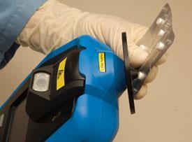
ChemImage, in the US, has developed a wide field hyperspeed imaging platform (Whip) technology, which can produce the first real-time Raman movies. And out of the lab, Ahura Scientific, also in the US, has introduced hand-held Raman spectrometers for security, military and forensic settings
Chiral clarity
For most structural biologists, x-ray crystallography and NMR make up the standard toolkit of techniques. But Blanch thinks differently. ’We are not able to get a pretty PDB [protein data bank] structure yet,’ he admits. ’But we can still get useful information from Raman that biologists and biochemists actually find important.’
Blanch’s team uses a technique known as Raman optical activity (ROA) to gain insights into chiral structural features of proteins and other biomolecules. ’Basically, ROA is the chiral form of Raman,’ says Blanch. ’In normal Raman you don’t really care what the polarisation state of the incident or scattered light is. But the polarisation state also contains a lot of information.’
Different chiral forms of a molecule scatter light with different polarisation, and also respond in different ways to circularly polarised light. So ROA can give rich insights into the structural folds and loops that define the function of biomolecules. Chiral features such as an alpha helix, a beta sheet, or a certain RNA fold can be easily recognised in the fingerprint.
Blanch has shown that ROA is a valuable tool for analysing unfolded proteins. He describes these macromolecules as ’nebulous, spaghetti type structures’ that are very difficult to crystallise so were often ignored by biologists. ’But they make up 20 to 40 per cent of our protein, so they’re doing something.’
Their unfolded nature makes these proteins flexible, and less specialised. They are thought to be employed by some enzymes to make them adaptable to a variety of substrates. ’ROA has proven useful because we can say more than just: that’s an unfolded structure. You can get different types of unfolded structure and we can look at the differences,’ says Blanch.
Another advantage of Raman techniques, according to Blanch, is that these studies can be performed in conditions that are close to ’real life’. Raman and ROA can be performed on proteins in solution, and can be used to monitor structural changes brought on by changes in pH or temperature.
One of the disordered structures Blanch has studied - polyproline II helix (PPII) - has been recognised as having an important role in protein folding. When this process goes wrong it can result in structures like amyloid plaques - the insoluble protein aggregates implicated in the development of diseases such as Alzheimer’s. ROA investigations have led Blanch and his colleagues to suggest that PPII has a critical, intermediate role in these formation processes.
’Other people are looking at the next step, which is to use Raman and IR [infrared] as in situ diagnostic probes,’ says Blanch. Meanwhile, advances like these in the understanding of the mechanisms behind Alzheimer’s onset could help with development of inhibitors.
Looking further ahead, Goodacre predicts Raman use will grow in the pharmaceutical sector as tighter regulations over metabolic manufacturing of natural products come into force. Raman fits the bill for monitoring these fermentation processes as it is a non-invasive technique which doesn’t suffer from water interference, unlike its rival near-IR.
Zenobi, meanwhile, is confident that single molecule TERS will be ’hugely successful’ in nanoscience, particularly since nanostructures such as carbon nanotubes have strong Raman signals. ’People are making nanowires out of carbon nanotubes and complicated systems from single molecules, but what you really have on the surface is not well known,’ he says. Zenobi also believes it will be possible to observe reactions on catalytic metal surfaces, using TERS to identify reacting chemical species. ’We have started to study this. If we could do it, I’d be getting a call from Sweden.’
According to Blanch, the basic tools needed to realise Raman’s potential are already in place. Some technological development is needed before Raman scanners are seen at airports and in hospitals, but more important is improving our understanding of what it takes to get a reliable result and understand the data. ’That should make all these methods more useful to biologists and chemists, government regulators and the rest of us,’ he says.
Tom Westgate is a science writer based in Manchester, UK
Further Reading
Faraday Discuss., 2006, 132, 3 (DOI: 10.1039/b600675m)
W H Zhang et al, J. Phys. Chem. C, 2007, 111, 1733 (DOI: 10.1021/jp064740r)
Also of interest
Surface enhanced Raman scattering
Chem Soc Rev issue 5 is a thematic issue on the topic of surface enhanced Raman scattering


No comments yet