
Rachel Brazil goes behind the scenes of molecular film sets
Chemists have devised many ways to illustrate molecules as they react, using everything from elemental symbols and curly arrows to 3D models. It’s easy to think these representations are reality – but how we picture reactions proceeding is usually based on kinetic or thermodynamic evidence. Do we really know what’s happening at the atomic level? What would we see if we could actually watch molecules reacting in real time?
Electron microscopes already allow us to dive deep into the molecular world. The 2017 Nobel prize in chemistry, for example, was awarded for advances in cryo-electron microscopy that allow protein structures to be seen at angstrom resolution. But static images of molecules are one thing – what about using electron microscopes to watch chemistry in action?
Molecular snapshots
To achieve this, we need to be able to image how the atoms in molecules re-arrange during a chemical process. The development of scanning tunnelling microscopy (STM), at the IBM research centre in Zurich, Switzerland, in 1981, was an important step towards high resolution imaging of molecules. A voltage applied between a surface and a very fine metal tip creates a tunnelling current whose size is dependent on the surface electron density. But the resolution isn’t high enough to see a molecule in detail – the electron density from the constituent atoms merge into one.
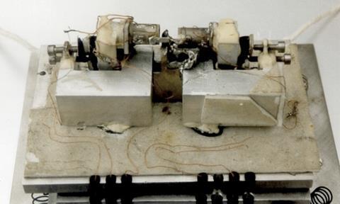
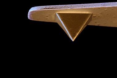
Then came atomic force microscopy (AFM), also from IBM, which measures the small force between the surface and the tip. For imaging, it is usually used in the non-contact mode where the tip is vibrated at a constant height above the surface. IBM Zurich researcher Leo Gross explains: ‘[The tip] is like a guitar string, it oscillates at its resonant frequency (about 30kHz) and this gets a little bit detuned due to the [surface] interaction and can be a little bit faster or a little bit slower than the resonance frequency, so what you see is always a frequency shift.’ Simply put, brightness in the image represents greater repulsive forces caused by greater electron density.
For almost the first time, you could actually image the bonds
Alexander Riss, TU Munich
As well as lowering the temperature and using ultra-high vacuum conditions, the key innovation that allowed scientists to really see molecular detail was the functionalisation of the AFM tip. The metal atoms in an AFM tip are bigger than the atoms in the organic molecules being imaged, and exert large attractive forces. Gross and his IBM colleagues found that a carbon monoxide molecule, picked up from CO deposits on the surface, absorbs onto the tip with the oxygen pointing towards the surface. As CO is inert, the tip could now get very close to the molecule being imaged without bonding with or dislodging them. ‘If there is some high electron density [the CO molecule] tilts away and this gives rise to image enhancement,’ says Gross. This effect seems to be linked to electron orbital repulsions between the CO electrons and the surface electrons, which increases the observed signal intensity.
‘For almost the first time, you could actually image the bonds and get a resolved structure of molecules and this was especially exciting for chemistry,’ explains physicist Alexander Riss at the Technical University of Munich in Germany. The images produced are remarkable and often show distinct single, double or triple bonds.
Reactions caught on camera
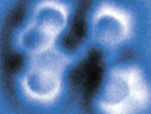
In 2013, a collaboration led by physicist Michael Crommie and chemist Felix Fischer, both at the University of California, Berkeley, US, reported an early effort to study chemical reactions using the IBM method. Riss was a member of Crommie’s research group at that time. One reaction observed was the chemical rearrangement of an enediyne – phenylene-1,2-ethynylene – on a silver surface.1 Images were taken before and after heating the molecule above 90°C. Three different products from cyclical rearrangements were revealed. ‘There is the formation of rings in there, there are some hydrogen transfers in there and all kinds of transformations, and it is experimental proof of the different mechanism possible from reactant to product,’ says Riss.
We would never have thought this would work
Leo Gross, IBM
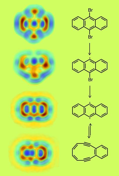
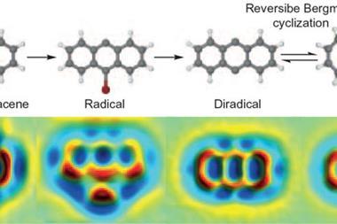
At IBM, Gross is also studying chemical reactions using AFM. Electrons from the tip are being used to initiate reactions; making and breaking bonds and shooting off atoms to form radicals. ‘Typically reactions where we shoot off hydrogen, iodine or bromine they need about two to three volts,’ Gross explains. In 2016, using a similar enediyne example, his team imaged each step in a molecule’s reversible isomerisation which was thought to occur via a mechanism known as the Bergman cyclisation.2
The 9,10-dibromoanthracene was stabilising on a sodium chloride bilayer which had been deposited on a copper surface. Electrons from the AFM tip first removed one bromine, then a second to form the diradical, and finally triggered the cyclisation. At each stage of the reaction process the molecule was taken back into the low-temperature, high-vacuum imaging conditions. The resulting series of images showed two radical intermediates – with one and both bromines removed – and the highly strained diyne with a fused six and 10-membered ring formed by the cyclisation. The reaction could also be watched in reverse – the first time that this had been achieved. ‘We didn’t expect that we could open and close the ring – we would never have thought this would work,’ says Gross.
Breaking or rearranging bonds is relatively easy, but ‘it’s much tougher to make bonds’, he explains. Other limitations of the technique include its preference for flat molecules. Topographical differences are difficult to differentiate from contrasts caused by different chemical bonding states. There is also an ongoing debate on the visibility of hydrogen atoms whose low electron density makes them near invisible.
But the biggest problem with using AFM to study reaction mechanisms is the inability to follow them live, from beginning to end without stopping to recreate the conditions needed for high-resolution imaging. ‘It’s very good in terms of spatial resolution, but very bad in terms of time resolution. We actually cannot look at the reactions while they are happening,’ admits Gross. ’You can only take snapshots before and after.’
Molecular film stars
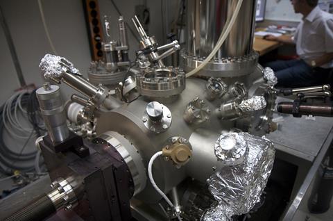
It is, however, possible to obtain stop-frames of molecules reacting using a transmission electron microscope (TEM). This technique, coined ChemTEM, is being developed by Andrei Khlobystov at the University of Nottingham in the UK. ‘We are imaging individual molecules and we can follow their fate through an entire reaction, from reactant via intermediate to product,’ he explains.
The technique uses a state-of-the-art TEM designed for sub-Ångstrom imaging, and takes advantage of what in most instances is a problem – sample damage caused by the electron beam. ‘ChemTEM turns the whole thing 180 degrees,’ explains Khlobystov. ‘It is not damage, it’s actually an induced chemical reaction, by virtue of the energy of the electron beam. We get the image of the reaction at the same time as we induce it.’
Currently, ChemTEM can image at a rate of about 0.5 to 1 seconds per frame and this limits the reactions it can observe. Khlobystov says, in principle, ChemTEM can go down to a 400 frames per second and he expects their capabilities to improve with the better instrumentation being developed by his collaborators at Ulm University in Germany.
We discovered something under the microscope which is completely astonishing
Andrei Khlobystov, University of Nottingham

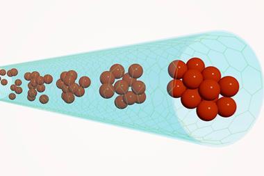
He has also found an alternative way to image faster reactions, using ChemTEM, by confining reactants inside a carbon nanotube. ‘The nanotube slows down the diffusion of molecules due to the confinement effect, it doesn’t allow them to move too far or too fast and the dynamics of the reaction decelerates, which is advantageous for imaging,’ says Khlobystov. As with AFM, hydrogens are still difficult to see using ChemTEM.
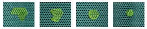
‘On a regular basis we discovered something under the microscope which is completely astonishing and surprising,’ says Khlobystov. An early surprise in 2010 came from imaging the mechanism of Buckminsterfullerene formation.3 For the previous 25 years it was thought impossible for fullerenes to form from a graphene sheet, they were believed to only form by assembling smaller carbon chains, first into rings and then fullerenes. Khlobystov’s ChemTEM proved this wrong. ‘You can image step-by-step the entire process of graphene transformation to fullerene – it is possible, we could show it directly in real space, at the atomic level and that was a big surprise,’ he says.
Another surprise captured on film was the formation of metastable intermediates in the condensation reaction of the disk-like pechlorocoronene molecule.4 The TEM electron beam removed two chlorine atoms on one side of the polyaromatic molecule creating an intermediate aryne (C24Cl10) biradical. ‘You could see it quite clearly dancing on the [graphene] surface for quite an extended period of time,’ says Khlobystov. In the nanotube, the aryne and a next-door molecule then undergo a Diels–Alder reaction to form a V-shaped intermediate, which then rearranges to release chlorine. The condensation could be repeated to create a long nanoribbon inside the nanotube.
ChemTEM could become a new tool for synthetic chemists, says Khlobystov. ‘TEM can shift the paradigm of how we discover materials, how we discover new chemical reactions. Not many new classes of reactions are reported every year, but I think there are more [to discover] in terms of what is possible in principle, and the only way to do it is to look at the matter at an atomic level.’
Having a gas
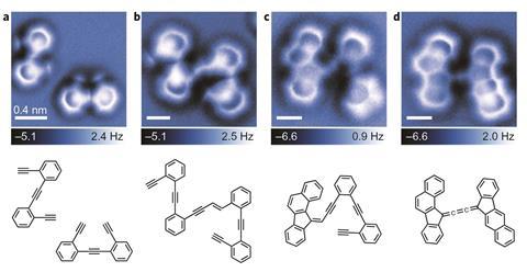
There are questions, however, about whether reaction pathways seen under the microscope are a true representation of how reactions proceed in the gas phase or in solution. Riss and his colleagues have demonstrated this issue using another enediyne cyclisation reaction.5 The reaction between two 1,2-bis(2-ethynylphenyl)ethyne molecules was imaged at low temperature on a silver surface, before and after heating.

Several products formed by a variety of cyclisation processes were captured, but the existence of some of these were surprising because they suggested mechanisms that seemed unlikely using theoretical models. Riss concluded that absorption on the silver surface stabilised some intermediates. ‘Some of the chemical energy is dissipated to the substrate and is then not available to the molecule anymore,’ he explains.
So is there a way to obtain the types of images or films we can now achieve for adsorbed molecules for gas or liquid phase reactions? Not yet, but ultrafast laser sources can help us peek at gas phase reactions in their early stages. In 2016, Jens Biegert from the Institute of Photonic Sciences in Barcelona, Spain, and collaborators used a form of laser-induced electron diffraction (LIED) to record the first direct visualisation of bond breakage in acetylene.6
Developed over the past decade, Biegert describes their method as taking a ‘molecular selfie’. His state-of-the-art ultrafast mid-infrared (IR) laser source is combined with a reaction microscope – a momentum spectrometer able to detect the 3D momentum and position of electrons and ions as they fly apart. An IR pulse removes an electron from the molecule and accelerates it back. On its return trajectory it kicks out another electron and is then scattered by the parent [C2H2]2+ ion – all within 9 femtoseconds. ‘If you just use one electron and you scatter it on one molecule you get the ultimate time resolution because the laser field dictates exactly when this electron hits,’ explains Biegert.
For the first time we catch a molecule as one of its bonds breaks
Jens Biegert, Institute of Photonic Sciences
The measurements are repeated around five billion times (which takes about a day) to get a statistical distribution and from this data, the various possible reaction outcomes are analysed including the carbon–hydrogen bond breakage. ‘Think about playing pool or snooker, if you collect where all the balls go and you know their direction and speed, you know everything about them. You can exactly calculate the structure that they had before you hit them, just like in particle physics,’ says Biegert. They were even able to detect the position of the hydrogen atoms.
Within a very small time window after the electron collision at 9fs, they were able to reconstruct the positions of the individual atoms in the acetylene molecule with a spatial resolution of 0.05Å and created several snapshots of the molecule. ‘For the first time we catch a molecule as one of its bonds breaks and a proton departs,’ says Biegert. ‘We are clear we see that the bond is broken because the proton is twice the original bond distance away.’
They detected various outcomes and in some instances a transient state was captured where the departing hydrogen atom and the remaining molecule fragment were still coherent, although the hydrogen had started to leave. The counter ion also shows evidence of isomerism, with extra hydrogen motion from one side of the molecule to the other. Currently the data hasn’t been converted into animations showing what is actually happening over time, but Biegert hopes to do so. ‘It would be extremely cool if we could make a movie of how this thing twists and changes its structure,’ he enthuses.
Biegert is now looking at more complex molecules like azobenze and hopes to image its isomerisation. Another ambition is to look at bond forming reactions. ‘Nobody, as far as I know, has really made bonds [using LIED] at least not in a convincing way,’ he explains. ‘The problem is that you need to bring two individual species very close together so they can interact and it’s very difficult.’
Seeing is believing
Jens Biegert
While their results do not represent the measurement of a single molecule reacting, Biegert’s team are able to probe chemical reactions at a femtosecond time scale – something currently not possible using STM or TEM.
For chemists, seeing AFM and TEM images so evocative of chemical stick diagrams is inspiring. But whilst these techniques may prove useful to us by allowing the study of surface catalysis, generally how chemically useful they will be remains to be seen. Could we one day be able to image complex chemical reactions or perhaps even biological processes at the atomic scale? There have been attempts to image DNA molecules at high resolution, but it hasn’t been possible to resolve individual bases yet.
Khlobystov is convinced that imaging reactions could create real advances in chemistry. ‘One day I want to see a TEM sitting next to NMR spectrometers and mass spectrometers as part of a normal chemistry laboratory,’ he says. But he does face scepticism: ‘I struggle to convince colleagues from organic synthesis that what I am doing has a chemical meaning.’ Nevertheless, he imagines that one day chemistry textbooks will replace stick diagrams with real images of molecules at the atomic level. ‘Students won’t have to believe the professor, they will watch reactions themselves – that would be wonderful.’ Biegert agrees: ‘It’s a bit cliché, but seeing is believing.’
Rachel Brazil is a science writer based in London, UK
References
1 D de Oteyza et al, Science, 2013, 340, 1434 (DOI: 10.1126/science.1238187)
2 B Schuler et al, Nat. Chem., 2016, 8, 220 (DOI: 10.1038/nchem.2438)
3 A Chuvilin et al, Nat. Chem., 2010, 2, 450 (DOI: 10.1038/nchem.644)
4 T Chamberlain et al, ACS Nano, 2017, 11, 2509 (DOI: 10.1021/acsnano.6b08228)
5 A Riss et al, Nat. Chem., 2016, 8, 678 (DOI: 10.1038/nchem.2506)
6 B Wolter et al, Science, 2016, 354, 308 (DOI: 10.1126/science.aah3429)


No comments yet