Neurodazine turns muscle cells from the sole of a human foot into something akin to stem cells
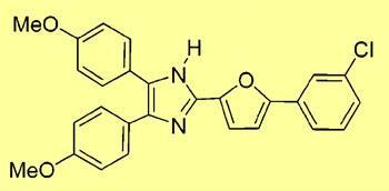
South Korean chemists say they have turned muscle cells from the sole of a human foot into something akin to stem cells, using a simple molecule called neurodazine.
If confirmed, their findings would be revolutionary, potentially allowing the development of new therapies for degenerative conditions like Parkinson’s disease without the ethical problems associated with using discarded human embryos to extract embryonic stem cells.
But other scientists are sceptical about claims that their method can convert fully-differentiated adult muscle cells into nerve cells.
Injae Shin’s team at Yonsei University, Seoul, cultured both mouse and human muscle cells in media containing neurodazine and other imidazole derivates. This group of compounds has a range of different biological effects and are used commercially as antifungal agents.
After a week, up to 50 per cent of cells in the culture showed changes in their structure and function indicating neurogenesis, or conversion into nerve cells. The cells developed the spindly growths (or neurites) typical of nerve cells and showed staining with antibodies raised specifically against various nerve cell proteins. In addition, these cells took up fluorescent dyes through a process linked to the depolarisation that occurs in activated nerve cells.
Cell doubt
However, stem cell biologists cast doubt on the team’s claims that they have produced a new source of stem cells. ’There is no convincing evidence for true neuronal differentiation in this paper,’ said Sally Lowell, head of the embryonic stem cell differentiation group at the Institute for Stem Cell Research, Edinburgh. ’A common problem is that many antibodies used to detect neuronal markers can also stain non-neuronal cell types under certain conditions,’ she said. ’The structures described as "neurites" could be confused with the short protrusions that are often seen extending from fibroblasts and other non-neuronal cell types.’

Shin accepts that other non-neuronal cells such as glial cells, which support and protect nerve cells, may develop similar shapes to those seen in the study but says Western blot analysis of antibody markers rules out this possibility. Moreover, he told Chemistry World, DNA chip data confirms that many of the genes involved in neurogenesis are activated in cells treated with neurodazine.
Yet David Tomlinson, a pharmacologist at the University of Manchester, was concerned about the apparent lack of proper controls in the study, to show that the markers used were not found in untreated cells. ’This is important because antibodies can bind non-specifically. Also there should be a demonstration that the cells are producing the messenger RNA for these markers,’ he said.
Location, location, location
Several scientists contacted by Chemistry World were also surprised by where the study was published. ’Real biological breakthroughs are not short communications in the Journal of the American Chemical Society,’ sniffed Gerd Kempermann, head of the Centre for Regenerative Medicine, Dresden, Germany.
However, Kempermann accepts that while it’s too early to be sure, the paper may yet prove to have scientific merit: ’It might well be that they are up to something interesting.’
John Bonner
Enjoy this story? Spread the word using the ’tools’ menu on the left.
References
et al J. Am. Chem. Soc. 2007, 129, 9258


No comments yet