Combined x-ray and laser technique explores reaction with light lasting trillionths of a second
Right now, light is damaging DNA in your cells, but the chemistry that causes this damage remains murky. But a team led by University College Dublin’s Susan Quinn has now tracked the precise dynamics behind the very first electron transfer in a key DNA damage process.
Exploiting the UK’s leading x-ray and laser facilities, Quinn and collaborators from the UK and Ireland studied periods lasting a few trillionths of a second, or picoseconds, and longer. ‘That single electron event is really important because free radical species form that can lead to mutation and therefore cancer,’ Quinn explains.
Such DNA damage happens commonly enough that we’ve evolved biochemical mechanisms to repair it, the study of which received this year’s chemistry Nobel prize. Yet we lack details about how light causes damage. ‘The first electron transfer steps are still not well defined,’ comments Jim Thomas from the University of Sheffield, UK, who wasn’t involved in the new study. We do know that guanine bases are particularly prone to oxidation though, adds Thomas.
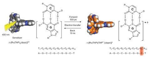
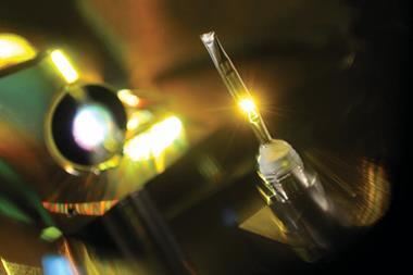
Together with John Kelly from Trinity College Dublin, Quinn has spent several years using spectroscopy to follow the very first stages of reactions involving light and DNA. They’ve studied DNA bound to ruthenium polypyridyl sensitiser complexes that make guanine oxidation happen even more readily in solution. Quinn and Kelly use time-resolved infrared (TRIR) spectroscopy at the Central Laser Facility (CLF) at the Rutherford Appleton Laboratory in Oxford, UK. There, they start the reaction with an ultraviolet laser and then an infinitesimally short time later investigate its status using an infrared laser. But with molecules jostling around in solution they’d been unable to determine precise locations for atoms participating in the reaction.
Diamond quality
The opportunity to remedy this emerged from the Diamond synchrotron, which is on the same site as the CLF. Using Diamond’s high-quality x-ray beams, Christine Cardin’s University of Reading team succeeded in the great challenge of crystallising DNA bound to ruthenium polypyridyl complexes. This meant the Dublin and Reading scientists could use TRIR spectroscopy to build on high-resolution x-ray crystallography atomic structure data.
This approach brings a new set of challenges, including demanding many more crystals than is typical for x-ray crystallography. The scientists sandwiched hundreds of 2.5–5µm crystal fragments, produced by James Hall from Cardin’s Reading group, between two transparent plates. Then, when performing TRIR spectroscopy, Quinn relied on Hall to confirm that she didn’t ‘fry’ the DNA. ‘The sample has to be in continuous motion, and the laser isn’t tightly focused, which ensures no single crystal is getting the entire pulse of energy,’ Quinn explains.
CLF’s facilities enable measurements over the picosecond to microsecond time range, in which period the researchers first tracked light exciting the ruthenium complex. They then followed the oxidation process transferring an electron from guanine to the excited complex, and the slower reverse transition, with the electron returning to the guanine. They were also able to spot precisely which guanine out of four in their DNA chain was reacting.
Tony Vlcek, a photochemistry expert at Queen Mary University of London, calls the result ‘truly remarkable’. ‘This work demonstrates the power of combined x-ray structural and time-resolved spectroscopic studies to investigate biological electron transfer,’ he says. ‘The physiological relevance of such studies, however, would vary from case to case, depending on structural and solvation dynamics.’
Thomas agrees that the combination of crystallography and TRIR spectroscopy is ‘clever’. ‘Because electron transfer is a key step in a wide range of biological processes, from cellular metabolism to photosynthesis, this new combined technique could offer real insights into fundamentally important questions in biology,’ he emphasises.
Among many potential follow-up studies, the team now hopes to use this approach to study native DNA. These results are also important because ruthenium complexes could eventually be used as light-activated chemotherapy, Quinn adds.
References
J P Hall et al, Nat. Chem., 2015, DOI: 10.1038/nchem.2369


No comments yet