A disposable polymer microchip promises to make medical diagnostics easier and more convenient.
A disposable polymer microchip promises to make medical diagnostics easier and more convenient, say Jo?l Rossier and Fr?d?ric Reymond.
Immunoassay is the workhorse tool of biomedical diagnostics. Annual sales for immunoassay reagents and supplies are currently ca $7000m (>?4000m) worldwide and ca $2000m in the US. In medicine, immunoassay is used in two general classes of applications: for identifying the organism responsible for a disease (diagnosis), and for monitoring disease treatment. Despite its success, however, immunoassay does have drawbacks. In particular long assay times, complex and expensive equipment, and the need for trained technicians have restricted its use mainly to centralised laboratories. But things look set to change. In this competitive market-place, medical diagnostic companies are already looking at new technologies that allow fast, quantitative and portable assays with simplified instrumentation.
Lab-on-a-chip systems capable of analysing minute volumes of sample currently offer the best hope. Microchips should not only greatly simplify the process of immunoassay, but could also allow diagnosis to be carried out in local analytical laboratories, A&E departments, doctors’ surgeries or even pharmacies. In this way, they may one day be used by all health professionals at ’point of care’ regardless of location, and ultimately even by patients in the home.
At the Laboratoire of Electrochimie of the Ecole Polytechnique F?d?rale de Lausanne in Switzerland, we first became interested in the area of microanalysis in the mid-1990s. Other researchers had at that time begun to report the first experiments performed on miniaturised platforms such as silicon chips. A few of these research groups had also demonstrated machining processes for fabricating polymer microstructures, though well-defined and reproducible manufacturing processes were still lacking.
Coming from the glucose sensing area, our laboratory head, Hubert Girault, had in mind the idea of developing new types of electrochemical biosensors. Electrochemical sensors have to date mainly been devoted to glucose sensing, which is one of the biggest markets for in vitro diagnostics. Our own interest, however, lay in developing a portable affinity system using disposable microchips for detecting proteins. We reasoned that we should be able to detect the interactions of proteins of interest with the corresponding enzymes or receptors by following small changes in electrical currents.
With this idea in mind, polymers (rather than silicon typically used for chip manufacture) seemed to be a good material of choice for our chips. Not only do proteins adhere easily to polymers, but for the purposes of electrochemical sensing our chip also needed to be non-conducting. As well as offering considerable flexibility, both in terms of performance and ease of processing, polymers have the advantage of being cheap and readily disposable - thereby avoiding any possibility of cross-contaminating samples.
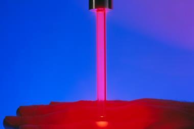
As a first step, we wanted to demonstrate the feasibility of polymer-based microsystems for generating electroosmotic flow and performing electrophoretic separations. By electrically controlling the flow of solutions along microchannels etched into the surface of a polymer chip, we believed that it should be possible to separate the various solutes electrochemically. Since our laboratory was already equipped with an excimer laser capable of drilling micro-holes in polymer sheets, we used photoablation to produce the microchannels by vaporising polymer, and then sealed these by lamination. The success of our electrophoretic separation depended on generating sufficient charges on the microchannel surface - and hence a big enough ’zeta potential’ - to control fluid flow.
With this achieved, the next step was to develop these microchips as electrochemical sensors, notably for protein assays. By 1997, we could integrate carbon ink microelectrodes within our microchips and use them as transducers - capable of detecting electrical currents resulting from, for example, an oxidation reaction. Then in 1998 we were able to use our chips to immobilise on the microchannel walls a radioactively labelled bacterial toxin, namely Staphylococcal enterotoxin B (SEB). Measuring the resulting levels of radioactivity confirmed the efficiency of our immobilisation procedure - an essential prerequisite for performing enzyme-linked immunoassays (Elisa). We went on to demonstrate the ability of the chips to perform such Elisa tests in 1999, in this case the test involved detecting a particular compound, D-Dimer, by following electrochemical changes.
To carry out such an immunoassay, we first immobilise on the walls of the microchannel an antibody that can specifically bind to an antigen of interest ( see below). Next, we allow a sample of plasma, blood or urine containing this antigen ( eg D-Dimer) to flow through the system. This antigen binds to the immobilised antibodies to form an antigen-antibody complex and excess sample is washed away. At this stage we introduce a second antibody labelled with an enzyme ( eg alkaline phosphatase, peroxidase or glucose oxidase) into the microchannel. This secondary antibody further binds to the antigen-antibody complex, and after washing away any excess, we add an enzyme substrate ( p-aminophenyl phosphate in the case of alkaline phosphatase) to the microchannel.
Immunoassay on a polymer microchip: chip preparation and assay procedure

The enzyme transforms this substrate into an electroactive product that can be oxidised at the electrodes, releasing electrons that are detected as an electrical current in the process. The magnitude of the current is therefore directly proportional to the concentration of the antigen or other analyte. This is a big advantage of electrochemistry, because the measured current is not limited by the number of analyte molecules present in the system but by its concentration; the response obtained with microchips is therefore identical to that obtained for much larger scale assays.
Our initial experiment worked well, but would our microchips be sensitive enough to compete with more conventional immunoassay techniques? To find out, we performed Elisa-type assays, with eg a biotin-alkaline phosphatase (ALP) complex in a buffer solution, to evaluate the detection limit and the dynamic range of the assay. After complex formation of biotin with avidin coated on the microchannel walls, we added an enzyme substrate, p-aminophenyl phosphate, to the avidin-biotin-ALP complex and this was transformed by ALP into p-aminophenol (Scheme 1).
Scheme 1. Electrochemical detection for Elisa assays

This p-aminophenol in turn can then be oxidised to quinonimide by applying a small voltage, releasing two electrons for detection. Our results showed that we could detect biotin down to subpicomolar (<10-12 mol l-1) levels - which compares very favourably with other immunoassay detection levels (which are typically in the range of 10-13 mol l-1 to 10-6 mol l-1).
On the basis of these early promising results, in 1999, we (with lab head H. Girault) officially launched DiagnoSwiss to develop and commercialise our microchips. DiagnoSwiss headquarters are in Monthey, close to both Lausanne and Geneva. The company started with just one R&D contract and has now grown to six employees.
One of the first steps towards commercialising our chips was to identify a technology for producing them in mass, and at low cost. Laser photoablation is a good prototyping tool, because you can use the laser as a pen to ’draw’ the structure you want to test. However, as a serial process, this is probably too expensive for large-scale production.
It was through our collaborations with a printed circuit board manufacturer that we hit upon the idea of adapting plasma etching technology to produce our chips. Plasma etching has the advantage of operating in parallel, which significantly reduces production costs. Most usefully, this technique allows us to integrate the electrode components of our chip directly, without the need for subsequent processing steps.
DiagnoSwiss chips are thin polymer foils made of polyimide and comprising microchannels with integrated gold microelectrodes. These chips are generic platforms that can be used for any micro-analytical application, but we are currently focusing on medical diagnostics and protein screening. Typically, our microchips consist of 50nl microchannels with a series of gold electrodes of 50?m in diameter, produced in 75?m thick polyimide films and sealed by a 35?m lamination layer of polyethylene (PE) covered by polyethylene terephthalate (PET). Our chips are not yet commercialised, but various laboratories already use them for research purposes.
One of the main advantages of our microchips is speed of analysis. Incubation times are generally less than five minutes, while detection takes under a minute. Conventional Elisa tests, by comparison, are typically done in well plates and take 1-2 hours for a quantitative result. The rate of detection in all these assays generally depends on how quickly the two binding partners diffuse together in the sample to form, say, an antigen-antibody complex. By reducing the volume of the sample, and hence the distance between molecules, complex formation occurs much faster. The large (microchip) surface to (sample) volume ratio - about 100 times larger than with standard well plates - also favours the immobilisation of one of the binding partners (eg the antibody) on the chip surface, accelerating antigen-antibody complex formation still further, and increasing the dynamic range of the assay.
Looking ahead
Our current prototype microchips allow for eight tests to be performed simultaneously. Because the entire analysis takes less than 10 minutes (including incubation, detection and washing), about 50 immunoassays per hour can be conducted on a routine basis. However, our equipment is still at the prototype phase, and at least half of the analysis time is currently for repeated manipulations. With a fully automated set-up, and by increasing the number of simultaneous tests, we anticipate that the output could be easily increased to over 200 tests per hour.
Ultimately, we envisage that the doctor/nurse would take a drop of blood from the patient while in the consulting room, deposit it on the microchip and push a button that begins the fully automated analysis. Conventional immunoassays are performed in about five minutes in our platforms. For tests requiring very high sensitivity, sample accumulation procedures may require extending assay times up to 15 minutes.
Combining the advantages of disposable micro-analytical systems with electrochemical detection has opened up new areas for electrochemical biosensors. Our microchips allow for high-performance and rapid quantitative assays in an easy-to-use and cost effective platform. With its electrochemical microchips, DiagnoSwiss also offers standardised tests that can be used in doctors’ offices as well as in centralised biochemical laboratories, or one day perhaps even by patients themselves.
Source: Chemistry in Britain
Acknowledgements
Jo?l Rossier and Fr?d?ric Reymond are chief scientific officer and general manager at DiagnoSwiss
Further Reading
- J. S. Rossier, F. Reymond and P. E. Michel, Electrophoresis, 2002, 23, 858.
- J. S. Rossier et al, CHEManager, 2002, 3, 28.
- J. S. Rossier et al, Lab Chip, 2002, 2, 145.
- V. Gobry et al, Proteomics, 2002, 2, 405.
- J. S. Rossier and H. H. Girault, Lab Chip, 2001, 1, 153.
- J. S. Rossier et al, Anal. Chem., 1999, 71, 4294.
- A. Schwarz et al in Proceedings of the µTAS ’98 workshop held in Banff, Canada, 13-16 October 1998, D. J. Harrson and A. van den Berg ( eds.), p 241. Dordrecht: Kluwer Academic, 1998.
1. Plasma etching: cheap as chips
Laser photoablation is a useful tool for producing prototype microchips because the laser can be used as a pen to etch the test structures. However, only one chip can be produced at a time, which makes this method of manufacture too expensive for large-scale production. As an alternative, plasma etching means that we can produce many microchips in parallel and reduce the production costs.
When we produce our prototype microchips using laser photoablation we expose a polymer foil to high energy UV laser irradiation which is shot through a mask that depicts the desired microstructure.

Plasma etching was originally developed by the semiconductor industry to produce printed circuit boards. However, we soon realised that it would work for our chip manufacture. We begin with a polyimide foil coated on both sides with a copper layer. This multilayer foil is coated with a photoresist, which is exposed to light in a high-resolution laser printer in order to define the desired mask pattern.
Chemical etching is then used to eliminate copper in these exposed areas, before exposing the foils to an oxygen plasma that will etch the polymer, resulting in grooves and holes in the plastic layer. The polymer foils are further exposed to plasma in order to form microelectrodes at the bottom of the grooves and the copper is removed by chemical etching. Electrically conductive tracks are maintained during this process in order to obtain electrical contacts, which are further coated with gold by electroplating. Finally, the microchannels are sealed by a second polymer layer.
2. Nanospray chips
In addition to medical diagnostics, our microchips can also be adapted to run alongside mass spectrometry (MS) analysis for pharmaceutical research including proteomics, drug profiling and clinical trials. Coupling disposable microscale devices with mass spectrometers is of great interest in the drug discovery process, and notably in functional genomics and proteomics.
For example, electrospray ionisation (ESI)-MS is now widely used for analysing complex biological molecules (Chem. Br., February 2003, p33). In its miniaturised nanospray version, the technique allows tiny (nl) volumes of sample to be measured with very low detection limits.
In ESI-MS the sample is squeezed through a capillary tube or needle and then ionised using an acidic or basic solution and an electric field to generate the spray. This creates highly charged droplets from which the solvent rapidly evaporates. Because of this evaporation, the droplets get smaller and are eventually drawn into the MS analyser. The main drawbacks of nanospray-MS for analysing peptides and proteins are the difficulties in handling standard nanospray needles, its lack of automation and the relatively long washing steps required to prevent cross-contamination.
To overcome some of these problems, DiagnoSwiss has recently extended its microchip technology to develop polymer nanospray chips that can be viewed as disposable needles for automated MS sampling. As well as being able to couple our chips to several different kinds of mass spectrometer, we have shown that they have good spray stability, easy on/off spray switching and good sensitivity and selectivity to multiply charged compounds. We are now working on developing the interface needed to support the nanospray tips in order to allow easy coupling with liquid chromatography systems, and to create precise alignment and accurate spray generation.
Contact and Further Information
Diagno Swiss
Rte de l’Ile-au-Bois 2, c/o Cimo SA, Case Postale, CH-1870 Monthey, Switzerland
Tel: +41(0)244714900
Fax: +41(0)244714901


No comments yet