The list of remarkable applications for graphene grows ever longer. This time, scientists in the US and Korea have shown that the single-atom thick carbon membrane can be used as a cover slip for an electron microscope to allow atomic-resolution observations of wet chemistry - something that is notoriously tricky to achieve.
The researchers wanted to investigate how platinum nanocrystals form from solution. 'Seeing the crystals form at atomic resolution allows us to understand and thereby exploit the precise growth process,' explains team member Alex Zettl, of the University of California at Berkeley, US. However, obtaining good resolution of liquid-phase processes with transmission electron microscopy is hampered by the difficulty of maintaining realistic conditions for the liquid specimen under the high vacuum conditions needed. One way around this is to enclose the sample in a sealed cell, with a window made from silicon nitride or silica. However, the lens needs to be relatively thick and so impedes the transmission of electrons, reducing the resolution that can be obtained.
Zettl and his colleagues figured that graphene might make for a good liquid cell. 'Graphene is continuous without holes, it is very flexible and very strong, and best of all it is only one atom thick, and it is very transparent to the electron beam,' Zettl says.
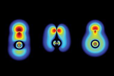
The team fabricated two layers of graphene and trapped a solution of platinum ions between them, creating a blister of graphene enclosing the solution. In the microscope, the incident electrons reduce the platinum ions to the metal, to form nanocrystals. The graphene liquid cell allowed the researchers to observe crystal growth in unprecedented detail. 'The formation shows more active faces attracting new atoms, and the merging of small crystallites into larger ones,' says Zettl. 'Different models have been proposed for the growth, but now we know which ones are correct by direct observation. It is a bit like seeing a finished ancient Egyptian temple and then proposing models of which blocks were put down in which order; but now we can watch the temple being built block by block. It gives us tremendous new insight.' The technique should be applicable to a wide range of liquid-phase systems, Zettl says: 'Basically, any wet chemistry.'
Valeria Nicolosi, who uses high spatial resolution electron microscopy to study two-dimensional nanomaterials, including graphene, at the University of Oxford, UK, is impressed by the work. 'I find this approach of using graphene as a vehicle for in situ liquid cell imaging revolutionary and innovative to say the least,' she says. 'They not only created an environment which realistically reproduces non-perturbative conditions for the liquid specimen, allowing in situ live investigation, but allowed atomic-resolution imaging and minimised heating effects under the electron beam.'


No comments yet