Identifying melanomas that have not metastasised could help patients avoid unnecessary and unpleasant additional treatment
Scientists in Belgium have shown that infrared spectrometry can help predict how likely it is that a melanoma tumour, the deadliest form of skin cancer, has spread to other organs.
Tumour spread, or metastasis, is the main threat to survival in melanoma patients. However, no biomarkers have been identified to indicate a high risk of metastasis and visual assessment of primary tumours by a trained pathologist is unlikely to reveal if cancerous cells have escaped to other parts of the body.
This is where the method devised by Erik Goormaghtigh and Noémie Wald at the Free University of Brussels could make a difference. Localised melanomas are treated very differently to those that have metastasised – surgical removal can be sufficient for localised tumours but metastatic melanomas require extra treatments like chemotherapy, radiotherapy and immunotherapy. ‘If it can be known for sure that a patient has no metastases, the doctor could avoid the ordeal of additional therapy,’ explains Wald. ‘Conversely, it is as important to identify those patients with skin tumours in a state of sending metastases that could appear benign under the classical microscope.’
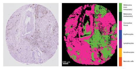
Goormaghtigh and Wald identified that primary melanoma tumours with and without metastases have different infrared spectra. Cells that become invasive acquire a series of biochemical characteristics that allow them to detach from the tissue and migrate through the blood and lymphatic circulation. Wald says these biochemical modifications are globally reflected in the infrared spectra. ‘As tissues are made from a huge number of molecules, it is not possible to assign the spectral differences to a single molecule. Yet, the infrared spectrum provides a very precise fingerprint of very faint differences between cells.’ These fingerprints were used to build a statistical model to automatically predict the invasive capability of tumours from their infrared spectrum.
Hugh J Byrne, head of the FOCAS Research Institute at the Dublin Institute of Technology in Ireland and an expert on the application of spectroscopy for diagnostics, thinks the study is an excellent example of how molecular based analytical techniques can be applied in a clinical setting. ‘Critically, the analysis can identify disease onset at the earliest stages, based on biochemical abnormalities, rather than morphological manifestations which are challenging to current clinical practice. The significant improvements in diagnostic capabilities offered by vibrational spectroscopy thus offer dramatic improvements for patient care and survival rates.’
Correction: On 13 January 2015 the story was updated as melanoma is not the most common form of skin cancer
References
This article is free to access until 24 February 2015. Download it here:
N Wald and E Goormaghtigh, Analyst, 2015, DOI: 10.1039/c4an01831a


No comments yet