New X-ray spectroscopy 'nanoreactor' watches catalysts in action
A promising technique for watching catalysts in action could provide new insights into how they work, report scientists in the Netherlands. The team used X-ray microscopy to study the catalytic Fischer-Tropsch reaction inside a specially-made reaction chamber, and say the technology should give scientists a greater understanding of catalytic activity, allowing better catalysts to be designed.
Solid catalysts are ubiquitous in the chemical industry, and accelerate the production of many important compounds. They are typically composed of nanometre-sized metal or metal oxide particles, attached to a solid support with a high surface area.
However, catalysts undergoes complex structural and chemical changes during reactions - so getting a direct look at the reacting catalyst can provide useful clues to improving their efficiency. But doing so at the temperatures and pressures commonly used in industry has so far proven difficult.
Now, a team led by Frank de Groot and Bert Weckhuysen at Utrecht University in the Netherlands, in collaboration with the Lawrence Berkeley National Laboratory, US, have achieved this using a tiny reaction chamber. The study represents the first time that a working heterogeneous catalyst has been examined at the nanoscale, the team say.
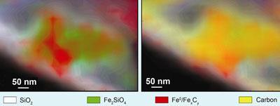
In De Groot’s ’nanoreactor’, reaction takes place between two windows etched to just 10nm thick. This setup allows X-rays to pass through the reaction and onto a detector, giving snapshots of the ongoing reaction. Some X-rays are absorbed by the catalyst, reactants and products - the exact energies absorbed revealing their chemical composition. The team were able to probe catalyst surfaces to a resolution of around 40 nm.
’This gives us a chance to examine the chemical changes that take place in a catalyst as it reacts,’ de Groot told Chemistry World, indicating that the resolution is sharp enough to pick out individual catalyst particles. ’This means it can provide us with a lot of useful information at the nanoscale.’
One reaction the team studied was the Fischer-Tropsch process - where a solid catalyst of iron oxide particles mounted on silica is used to convert carbon monoxide and hydrogen into liquid hydrocarbons that can be used as fuels.
De Groot and Weckhuysen’s team replicated the reaction in their nanoreactor and found that during the reaction the iron oxide underwent several transformations. The initial iron oxide (Fe2O3) is converted into another oxide (Fe3O4), before iron silicates (Fe2SiO4) and metallic iron begin to form. Finally, iron carbides (FexCy) begin to appear. By correlating the catalyst make-up in different areas with the organic products being formed, the team showed that carbon accumulates in the iron-rich areas, with the hydrocarbon products spilling from the metal onto the silicon support.
’These findings show great potential for heterogeneous catalysts to be imaged in situ,’ says Alexis Bell at the University of California, Berkeley, US. ’I imagine that the process is not limited to the observation of catalyst particles, but could also be used in many other applications, such as detecting levels of airborne particulates responsible for the formation of acid rain.’
Other proposed uses include monitoring structural changes in hydrogen storage materials or examining the distribution of medical nanoparticles in cells. The team say developments in optics and imaging methods should improve the technique’s resolution.
Lewis Brindley
Enjoy this story? Spread the word using the ’tools’ menu on the left.
References
E de Smit et al, Nature, 2008, DOI: 10.1038/nature07516


No comments yet