Real-time Sers monitoring of radiation treatment in a 3D prostate cancer model
Surface-enhanced Raman scattering (Sers) can help optimise radiotherapy treatment regimes for prostate cancer, UK scientists have shown.
Prostate cancer is the second most common cancer worldwide in men, and the fourth most common cancer overall. It is usually treated with radiotherapy, and doctors would like to tailor doses for individual patients. This would help maximise tumour cell destruction, while minimising damage to normal tissue.
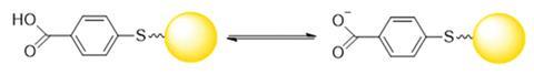
With this in mind, Colin Campbell and colleagues at the University of Edinburgh introduced gold nanoshells functionalised with pH and redox probes into prostate cancer cells, then measured their spectral fingerprint using Sers. Ionising radiation causes the cells’ mitochondria to leak their alkaline contents. This increases the pH and reduces the redox potential inside the cell, triggering cell death and changing the Sers fingerprint.
Using Sers probes also allows the team to examine 3D cell cultures in real time. Molecular methods to measure cell death require extensive sample preparation, and while traditional Raman spectroscopy can monitor live cultures, low signal intensity limits it to 2D cell monolayers. 3D spheroids provide a model that more closely resembles real tumours.
‘Measuring the physical chemistry of tumour spheroids, like changes in redox potential and pH, is undervalued in the community. Researchers typically look for changes in a particular protein or RNA,’ Campbell says. ‘The ability of Sers to do that in real time in a reversible and dynamic way makes it a valuable tool that gives you information you can’t get from other techniques.’
Using the Sers probes, the team showed that irradiating tumour spheroids with two daily doses of six gray (1Gy = 1J/kg of absorbed radiation energy) is more effective than a single dose of 12Gy, or thee daily doses of 4Gy.
‘The combination of nanoprobes specific to intracellular redox potential and pH provides a unique picture of the cellular response to radiation treatment,’ comments Zachary Schultz, an expert in vibrational spectroscopy at the University of Notre Dame in the US. ‘Another unique aspect is the extension to tumour mimics, which include more biological complexity than traditional cell culture. The design and analysis of these Sers nanoprobes and other nanomaterials has exciting possibilities for elucidating complex biological systems.’
Campbell suggests that because Sers is cheap and portable, with short acquisition times, it could eventually be used to screen cancer patients and personalise radiotherapy. The team intends to test the technique on more complex 3D spheroid models incorporating extracellular matrix components and other cancer cell types. The method could also be extended to predict chemotherapy drug efficacy and safety.
References
V Camus et al, Analyst, 2016, DOI: 10.1039/c6an01032f


No comments yet