Teams at Imperial College London prepare to focus on Alzheimers' disease and nanoscience of osteoporosis.
Tom Westgate/London, UK
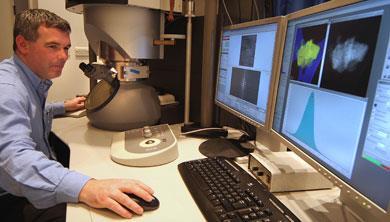
For the first time in the UK, researchers will be able to ’see’ atoms and the bonds between them, thanks to the country’s most powerful commercially available electron microscope. The London Centre for Nanotechnology, this week unveiled its brand new FEI Titan 80-300 monochromated scanning transmission electron microscope (STEM) at Imperial College, London, UK. It is the only Monochromated electron microscope in the UK, and there are only a handful in the world.
The Titan will be used in a range of projects, including an investigation of bone structure. ’It came as revelation to me that on the atomic scale, bone is not well understood,’ said David McComb, reader at the department of materials at Imperial, who has overseen the installation of the ?3 million facility. McComb hopes this will be ’a good starting point’ from which to understand how osteoporosis develops.
Another project will investigate the interface between brain tissue and iron nanoparticles which are formed in the brains of patients with Alzheimers’ disease.
The Titan can focus electrons to a probe of 0.14 nm. ’That’s pretty much on the scale of atomic resolution,’ said McComb. ’Many other instruments around the world will allow you to see on the atomic scale’ he added, but McComb is not satisfied with simply visualising atoms. ’Far more important these days is the identity of the atom that you’re seeing and more importantly how the atom is coordinated to the other atoms around it,’ he said. This close examination of interactions and interfaces is so useful ’because it’s how those atoms are actually bonded that will dictate the structure-property relationship.’ Very few instruments around the world can carry out this sort of analysis.
STEMs make an image by scanning the probe over a sample, and detecting the energy that is either scattered back from or passes through these tiny points on the sample. The energy loss data can be converted into an image which is related to the atomic number of the element: ’The heavier the element, the brighter the image,’ explained McComb. Bringing the imaging and the spectroscopy together in this way ’we can see the atom and ask: "what’s the chemistry? what’s the structure? what’s the bonding?"’ he said.
What makes this microscope stand out from the crowd is the monochromator. The monochromator does for electrons what a prism would do to white light. It splits the electron beam into the constituent wavelengths, researchers can then use a slit to select a particular wavelength and look at how that particular wavelength interacts with the sample. This improves the resolution of the spectrometer sufficiently that researchers can really see all of the changes associated with atoms whenever they change their environment, said McComb.
The microscope is so sensitive it required a special room to be built in the basement of the London Centre for Nanotechnology. The tiniest vibrations, temperature changes or electromagnetic fields had to be excluded from the facility - not easy in a Victorian building in central London. This task included identifying and insulating several original heating pipes which had electrical appliances earthed to them, and surrounding the room with copper coils. Current can be passed through the coils to compensate for any other electrical fields, and bring the net field in the room to zero, ’just to be on the safe side’ said McComb.
Putting the nano into nanochemistry
The discovery of a new form of elemental carbon 20 years ago changed thinking in chemistry. Philip Ball investigates whether the buckyball has lived up to the hype and what legacy ...
Pointy tip is just one atom wide


No comments yet