A new technique that colour-codes cancerous and healthy brain cells according to their chemistry could help surgeons remove all traces of brain tumours while minimising damage to sensitive tissues.
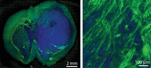
This is the latest in a series of developments to boost the chances of successful cancer surgery. Last month scientists unveiled a knife that analyses smoke released during surgery to detect whether tissue is healthy or cancerous. This technique, developed by Sunney Xie at Harvard University, US, and colleagues, uses stimulated Raman scattering (SRS) microscopy to detect lipids and proteins in brain tissue. Tumours are relatively high in protein and low in lipids, while normal brain tissue is rich in both. The team encoded the Raman signal for lipids in green and the signal for proteins in blue, so tumour cells appear blue and healthy ones green.
Using the technique on mice, the team were able to examine the margins of brain tumours and spot places where cancer cells have infiltrated healthy regions of the brain. These very fine details are impossible to see without SRS, but could help surgeons avoid removing too much or too little tissue. The team are now working on building a toothbrush-sized probe that could be used for continuous monitoring during surgery.
References
M Ji et al, Sci. Trans. Med., 2013, DOI: 10.1126/scitranslmed.3005954


No comments yet