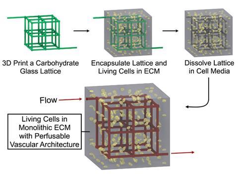
Now, US researchers can build vessels into a cell-containing gel – the beginnings of a thick tissue. Scientists form the gel around a lattice of printed sugar fibres. The fibres dissolve after the gel sets, leaving a network of channels that carry nutrients like blood vessels.
For the past decade, tissue engineers have looked for ways to build a 3D tissue in such a way that vessels are immediately available to feed growing cells. One way to create these vessels uses a tiny silicon template to pattern grooves in a sheet of cell-containing gel. Covering these cut outs with another sheet of engineered tissue creates enclosed channels. While these sheets can be layered to build up a tissue, the vessels only extend through the tissue in two dimensions, unlike the 3D network in our bodies.
Scientists can also print tissues using an inkjet printer to layer drops of cellular ink, leaving gaps for vessels. But they have to optimise the print settings for each cell type and supporting matrix.
Glassy sugar
Instead of printing delicate cells, Christopher Chen, of the University of Pennsylvania, and his colleagues used a 3D printer to create a sturdy vessel scaffold. They printed the scaffold fibres with a glassy sugar made of sucrose, glucose and dextrans. The structure was then covered in a solution of cells and molecules that tangle into a matrix to support the cells. Polymerising the matrix traps the cells in a gel and water dissolves the sugar fibres, leaving empty channels.
The growing tissue can then be fed by pumping nutrients through the channels. The scientists can also make these channels function more like natural blood vessels by lining the walls with cells. Because this new method works with a variety of cell types and water-based matrices, tissue designers have the freedom to choose the best matrix and cell combination for the tissue they want to grow, Chen says.
To grow an organ, scientists need to build a tissue with cells from that organ. However, liver cells are notoriously difficult to culture, says Abraham Stroock, of Cornell University, who was not involved with this study. Chen’s scaffolds kept more rat liver cells alive and functioning after eight days compared with those grown in a solid slab of gel.
Chen also built a vessel network and shipped it to other labs so those scientists could culture thick tissue from cells they harvested. In the future, you could imagine pre-printing vessels for hearts or other organs, Chen says. When doctors want to start growing an organ for transplant, they could pull out a sugar network, cover it with matrix mixture containing a patient’s cells and polymerise the matrix within 30 minutes, he adds.
The glassy sugar material used to print vessels that dissolve without damaging cells is the key advance of this work, Stroock says. But building vessels in a thick tissue doesn’t mean that all challenges of tissue engineering are solved. Stem cell scientists are still working to find the right cells to form specific tissues, Chen says.
Nevertheless, Jeffrey Borenstein, of Draper Laboratory, says Chen’s method is ‘a big step for tissue engineering’. While spun sugar fibres have been used to make blood vessels in block of silicone, this approach allows engineers to build a truly 3D vessel network in engineered tissue without damaging the cells. But, he adds, the printer may not be able to control the fibre diameter and shape to create the variety of branched vessels in natural vasculature.
Chen agrees that creating branching vessels with a range of diameters that mimic those in our bodies remains challenging. Jordan Miller, a postdoctoral student in Chen’s group, says the printer can create fibres thinner than a capillary, so their technique could be used to create more complex networks.
References
- J S Miller et al, Nat. Mater., 2012, DOI: 10.1038/NMAT3357


No comments yet