Tiny diamonds can be used to visualise cells and tissues and could deliver drugs too
A new microscopy technique for viewing ordinary industrial nanodiamonds in living cells has been unveiled by UK researchers. The researchers believe the technique could prove useful in medical research and treatment. Nanodiamonds are highly promising medical imaging contrast agents and drug delivery vehicles because of their low cytotoxicity. However, many applications being developed require nanodiamonds with fluorophores, such as nitrogen vacancy centres, which fluoresce upon laser excitation. These are expensive and difficult to produce in a controlled way.
Non-fluorescing nanodiamonds can be analysed using Raman scattering. The sample is bombarded with light, which interacts with the vibrating chemical bonds: the scattered photons reveal those bonds’ vibrational frequencies. However, conventional Raman scattering is weak, so producing a clear image takes a long time, which is problematic in a living organism. Coherent anti-Stokes Raman scattering (CARS) overcomes this problem with the use of two incident infrared lasers. When the difference between the frequencies of the two beams is equal to the vibrational frequency of the chemical bond, all identical bonds are driven to vibrate in phase, amplifying the signal and shortening the time it to takes to build up a picture. This is particularly beneficial for nanodiamonds, where a large number of identical carbon–carbon bonds are present.
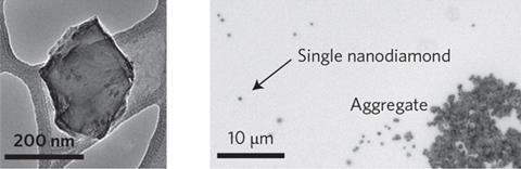
Paola Borri and colleagues at the University of Cardiff imaged nanodiamonds with radii of 70–150nm in water using a CARS microscope that they built themselves. Extrapolating from the images, the researchers calculate that they should be able to detect diamonds with radii as small as 27nm. Many medical nanoparticles are much smaller – sometimes as small as 1–2nm – but Borri believes the nanodiamonds could still be useful.
‘To target specific cells, for example cancer, it’s actually important that the nanoparticles are not too small so they will not be eliminated,’ she says. ‘Importantly, we can now relate the CARS signal strength with a quantitative measure of the nanodiamond size for single particles. This direct in situ determination of the nanodiamond size inside living cells is not possible with fluorescence techniques.’ The researchers achieved clear images of the diamonds in lab grown human cells, which were not damaged by the process.
Materials scientist Yury Gogotsi at Drexel University in the US is intrigued, although he sees a potential problem with using larger nanoparticles, with proportionally lower surface area, for drug delivery. ‘It may be that a therapeutically insignificant dosage can be delivered compared to, say, 5nm diamond particles,’ he says. Ultimately, he concludes, ‘It’s almost never obvious from the first results where the real applications will be.’


No comments yet