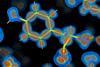
A 3d chemical protein structure with a heat map laid over.

Source: © Ashwin Chari/Max Planck Institute for Multidisciplinary Sciences & Gleb Bourenkov/European Molecu

The valence electron deformation map (blue to red: low to high density) of the small electron transfer protein rubredoxin at a resolution of 0.43 angstroms 

A new record has been achieved in protein imaging, capturing the protein rubredoxin at an unprecedented resolution of 0.43 angstroms, revealing structural features at a level rarely achieved for biological macromolecules.1

The measurement was made using x-rays from Petra III in Hamburg, Germany, one of the brightest synchrotron sources in the world. Previous experiments, in which the team explored how x-rays interact with and alter protein structures, as well as how radiation damage can affect collected data, allowed them to develop a protocol that minimises damage while collecting diffraction data from protein crystals.2

‘Strictly speaking, x-ray diffraction is not an imaging technique but … [we used] a highly uniform x-ray beam to illuminate crystals of biological macromolecules in multiple orientations,’ explains Ashwin Chari of Max Planck Institute for Multidisciplinary Sciences in Göttingen, Germany.

The team used a tailored x-ray beam with a uniform intensity known as a ‘top hat’ beam. Its size and shape can be adjusted to match each protein crystal, allowing precise control over the dose of x-ray radiation delivered during the experiment.

‘Reaching 0.43Å resolution for a protein is certainly an exceptional technical achievement,’ comments Anna Krawczuk at the Georg August University Göttingen who was not involved in the study. ‘The significance of this study goes beyond setting a new resolution record. At this level of data quality, we are no longer looking only at where the atoms are; we can begin to see how electrons are distributed around and between them. In other words, the experiment starts to provide information about the chemical bonds themselves.’

Crystallographic studies of small molecules have been used to probe their atomic and electronic properties since the 1990s. However, achieving the resolution and data quality needed to see these features in proteins has proved considerably more difficult.

‘This type of information has been accessible [through] crystallographic studies of much smaller molecules [for years], but obtaining data of sufficient quality for proteins is considerably more difficult,’ says Krawczuk. ‘The study therefore provides an important bridge between conventional structural biology and what is often called quantum crystallography, where the aim is not simply to determine molecular structure but also to extract information about its electronic structure.’

The approach could eventually open the door to routine quantum crystallography of biological macromolecules, says Chari, giving researchers deeper insight into how they function and control biological processes. This could include designing molecules that bind more precisely to biological targets and understanding how local electric fields drive enzymatic reactions.

Achieving this will require collecting many sub-angstrom structures of enzymes as they pass through different stages of a reaction. Chari believes this could be possible within the next decade.

Krawczuk says that there is still a considerable distance between what has been demonstrated here and obtaining a fully experimental picture of electron distribution in a full protein.

‘Although the experiment reveals features associated with the redistribution of electrons during chemical bonding, the detailed aspherical electron-density model used to describe these features is not itself determined entirely from the experimental data,’ she notes. ‘Another fundamental limitation is radiation damage. This is particularly important for biological materials … and cannot simply be eliminated. For future studies aimed at extracting increasingly subtle electronic information, understanding and controlling such effects will become even more critical.’

‘The next challenge is to understand how far these methods can be extended to more complex and less ideal biological systems,’ Chari says.