
Chemists have tracked the fleeting intermediates in the natural synthesis of penicillin using ultrashort x-ray pulses. The research could usher in improvements in ring-forming reactions on useful intermediates for the pharmaceutical industry or aid enzyme engineering.
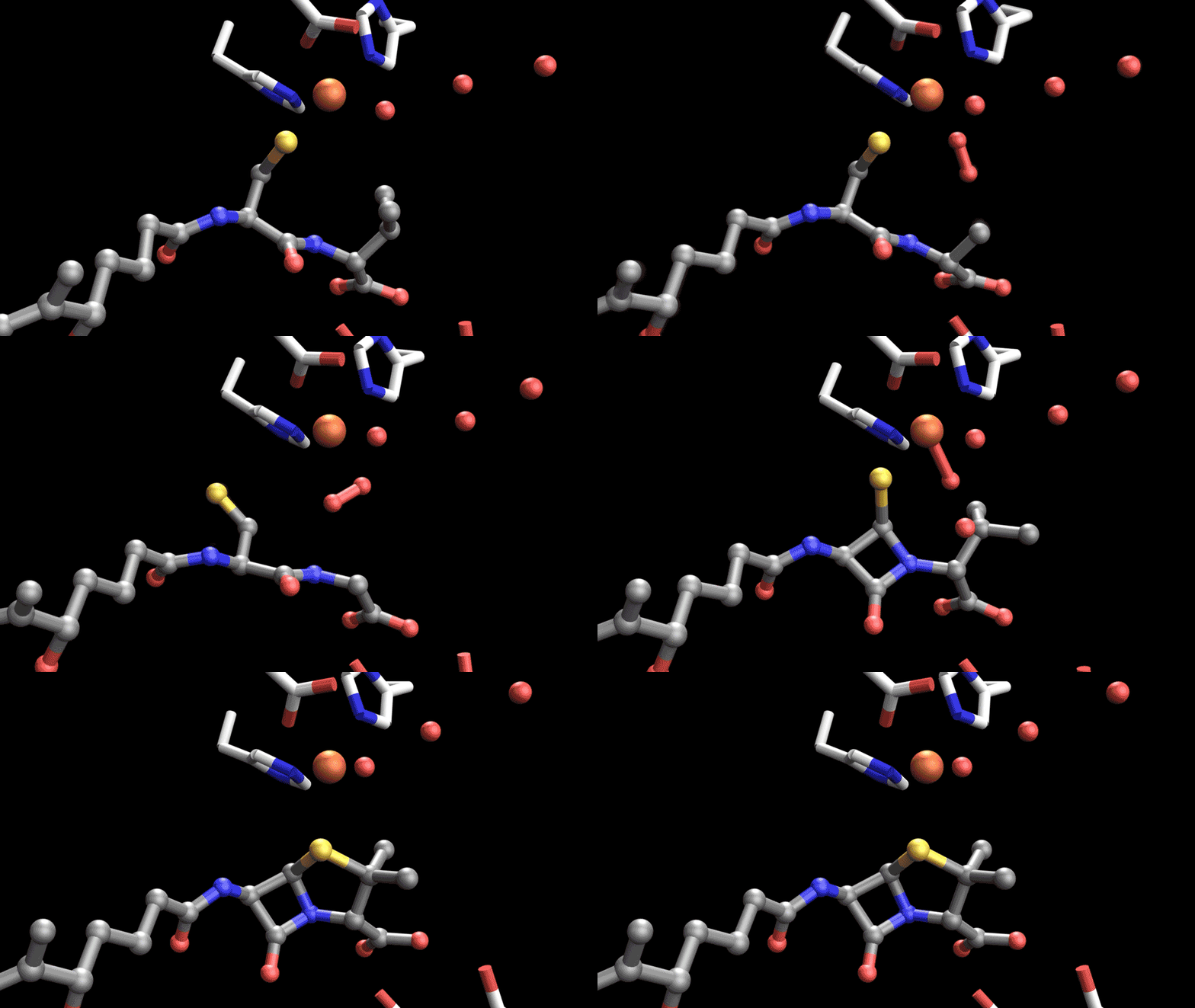
Penicillins and related ß-lactam antibiotics remain crucial medicines, accounting for more than half of the antibiotics given each year. Until now, the intermediates involved in the reaction have been too short-lived to observe directly and the complex ring-forming reaction has never been performed with a chemical catalyst.
‘Chemical synthesis of penicillins is very difficult and rather inefficient and we still don’t have the stereochemical control to achieve the right bioactive strained molecule,’ explains Christropher Schofield, who led the research at the University of Oxford.
The enzyme isopenicillin N synthase (IPNS) catalyses the conversion of a linear tripeptide into a fused four-membered ß-lactam and five-membered thiazolidine ring system characteristic of penicillins. The reaction requires breaking two relatively unreactive C–H bonds while creating the highly strained ß-lactam ring essential for antibiotic activity.
The international collaboration, which involved synchrotrons and x-ray free-electron lasers from the US, South Korean, Japan and the UK, followed the formation of penicillin using ultrafast x-ray pulses. A stop-motion reel of the reaction was created by depositing tiny droplets containing enzyme microcrystals onto a moving tape. The time-resolved structural snapshots from thousands of crystals were placed in sequence to create a ‘molecular movie’.
The enzyme was revealed to make use of molecular oxygen, which binds to ferrous iron to form an Fe(III)-superoxo species. This abstracts the hydrogen atom from the cysteine of the tripeptide substrate, leading to the formation of a thioaldehyde. Surprisingly, the thioaldehyde intermediate was not coordinated to the iron. The enzyme therefore stabilises and controls a highly reactive intermediate even though its sulfur is no longer directly attached to the catalytic metal.
‘The time resolved work now shows that a key intermediate transiently comes off the iron and then rejoins again [after ß-lactam ring formation],’ says Schofield. The iron–sulfur bond is restored as the monocyclic ß-lactam intermediate forms. ‘This was not predicted by high-level quantum mechanical calculations,’ says Scholfield.
Water molecules within the enzyme’s active site helps to guide the chemistry. ‘We think that water is somehow involved in proton delivery or electron acceptance,’ says Patrick Rabe, a chemical biologist with the Oxford team. ‘You get motions throughout the entire structure that exert conformational control,’ adds Schofield.
This water network around the active site is a striking result, notes David Sherman, a biological chemist at the University of Michigan. ‘Water molecules are not passive bystanders,’ he notes. ‘They are part of the catalytic machinery and the evidence supports their participation in proton transfers that help the first ring form.’ For enzyme engineering, ‘a particularly useful lesson is that residues outside the immediate iron-binding site deserve considerable attention. They can influence the chemistry through water positioning and electrostatics’.
‘This is the first time we can observe these intermediates for this class of enzyme. It is a tour-de-force piece of work,’ says chemist Greg Challis at the University of Warwick, UK. ‘The reaction pathway of this and related enzymes can be very complex and there’s lots of reactive, discrete intermediates that are transient. It is the holy grail of enzyme chemistry to be able to observe each of the successive intermediates in the pathway.’
References
P Rabe et al, Nat. Catal., 2026, DOI: 10.1038/s41929-026-01618-4






No comments yet