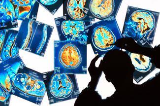
Boundaries of magnetic resonance imaging are continuing to be pushed to reveal more about the human body and aid disease diagnosis
Boundaries of magnetic resonance imaging are continuing to be pushed to reveal more about the human body and aid disease diagnosis
Imagine discovering when you are 40 that in 20 years you may develop Alzheimer’s disease symptoms. The doctor prescribes a change in diet and a more rigorous exercise regime, allowing you to stave off the devastating effects of the disease and live to a ripe age of 100 with all your faculties intact.
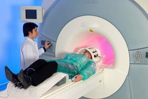
This vision is still fantasy, but using magnetic resonance imaging (MRI) - a technology that shows the internal anatomy of various body parts - researchers are turning it into reality. Unlike the widely used computed tomography (CT) scans, MRI doesn’t use x-rays and it provides greater contrast between the different soft tissues of the body making it particularly applicable for neurological, musculoskeletal, cardiovascular and oncology imaging.
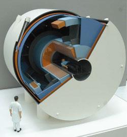
Its principles are the same as those of the chemist’s friend, nuclear magnetic resonance (NMR). MRI’s central component is a strong magnet, which generates a magnetic field that aligns the magnetisation of certain nuclei in the human body. Pulses of radiofrequency (RF) from detector coils in the machine introduce electromagnetic field gradients that cause the nuclei in the body to rotate and produce tiny magnetic fields for detection. By manipulating the rotating magnetic fields with various gradient coils, a detailed anatomical image of a body part - such as the brain, heart or knee - is created.
The field strength of a MRI instrument’s magnet is measured in units of tesla (T). When these machines were first built in the 1970s, the magnets were 0.1-0.5T. Since then their strength has grown incrementally, and today 1.5T and 3T magnets are common in clinical MRI laboratories.
But the research community are looking at stronger magnets still - 7T and upwards - to obtain better signal-to-noise (S/N) ratios. S/N increases approximately linearly with magnetic field strength, and researchers are greedy for more. ’S/N is like money. The more you have, the better,’ explains James Meng, director of Siemens Healthcare’s magnetic resonance division in Malvern, US. The extra signal can be used to get better spatial or temporal resolution in the images, or to image at a faster speed, he adds.
Super-sizing MRI
The 7T machines have turned out to be surprisingly useful, according to Bruce Rosen, director of the Athinoula A Martinos Center for Biomedical Imaging at the Massachusetts General Hospital, US. ’It’s surprising in the sense that people didn’t think the pictures were all that bad at 3T. But we’re discovering a whole series of features that were just at the limits of detectability at 3T that really stand out clearly at 7T.’
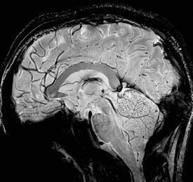
Alan Koretsky, an MRI expert at the US National Institute of Neurological Disorders and Stroke in Bethesda agrees. ’You start seeing things that have never been seen before in the brain,’ he explains. ’At 3T all white matter looks alike and all grey matter look alike.’ But at 7T, he says, the heterogeneity of white and grey matter in live brains can be visualised - something that was only previously seen in histological brain slices. Details, like lesions in the brain caused by strokes or spots responsible for epileptic seizures, can now be seen in live subjects.
A research team led by Rolf Gruetter at the Centre for BioMedical Imaging in Lausanne, Switzerland, is carrying out ground-breaking tests using high field machines. In one such experiment, the amount of oxygen in the blood in different areas of the brain was monitored while the fingertips of human subjects were stroked. ’We could see in the brain each single finger represented very precisely at 7T, something that’s hardly possible at lower magnetic fields,’ exclaims Gruetter. Magnetic susceptibility of the body tissue is heightened at the higher magnetic fields, he explains. ’You get exquisite contrast in the tissue, especially in the brain, that was previously not possible.’
Beyond hydrogen
Up to field strengths of 3T, researchers can easily track water protons in humans because the concentration of these protons is so high (80M) in the body. But as chemists know from NMR, nuclei such as sodium-23, phosphorus-31, carbon-13 and nitrogen-15 also give signals in a magnetic field. But unfortunately, their concentrations in the body are low. Sodium-23, an atom critical for biological function, is the next most-sensitive nucleus in a magnetic field after the proton but has only an average concentration in the body of 40mM. On top of that, because the sodium nucleus is heavier than a proton, it’s less sensitive to the RF electromagnetic fields and therefore harder to detect.
But with higher magnetic field machines, researchers are beginning to see signals from sodium-23 in humans. Keith Thulborn at the University of Illinois in Chicago, US, and his team have built a 9.4T MRI machine with which they can obtain high-quality images of the brain based on sodium ions.
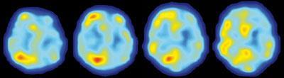
The brain is one of the most tightly packed tissues in the body, with an 80 per cent cell volume fraction. ’If you have any sort of disease that disrupts cell packing - for example, a tumour where you have invading neoplastic cells or stroke where you have cells dying - you change the cell density,’ says Thulborn. Using their high field instrument, the group have observed changes in cell-packing density by imaging the sodium ions in the cells.
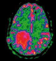
Another potential application for sodium ion imaging is Alzheimer’s disease. By the time the illness has clinically manifested, the brain is already being destroyed and current medical interventions are not potent enough to reverse the damage already done. But the actual pathological process is believed to start 20 to 30 years before the patient experiences any symptoms. The first tissue damage occurs in the hippocampus, where some cell types die causing a change in cell density in this area of the brain.
’Suppose we could use sodium imaging to detect the first cell loss in the hippocampus two decades before the patient was symptomatic? We would have the possibility of intervening on that disease process,’ envisions Thulborn.
Spectroscopy in an MRI machine
The better S/N ratios at high magnetic field strengths also lets researchers tackle NMR-type spectroscopic experiments in a MRI machine. Gruetter’s team has achieved biochemical analyses in vivo at 7T where they collect data from a live human brain, discard the signals from the water and fat protons, and focus on the signals coming from soluble metabolites in the cell. ’It’s an instantaneous assay of typically 20 compounds,’ he explains. Using the technique they have been able to track glucose uptake and transport in vivo to better understand energy transduction in cells.
Diabetes research is an area to potentially benefit from spectroscopy in high field MRI instruments, as being investigated by Dean Sherry’s team at the University of Texas at Dallas, US. The group is looking into how the movement of fat molecules out of the fat cells in skeletal muscle and into muscle cells spells trouble in diabetes. The investigators can pinpoint the locations of fats in the different cell types in live human subjects by looking at the peaks generated by the CH2 groups in the molecules. ’They have different chemical shifts,’ explains Sherry. ’You can actually resolve them by saying "here’s a peak that’s fat inside your muscle cell and here’s another peak that’s fat between your muscle cells". It’s really very powerful and an example of things you can do at high field.’
By tracking the location of the fats in the different cells of muscle, the investigators can start understanding the effects of exercise and diet on the progression of diabetes.
Jumping hurdles
The high magnetic field machines are not yet perfect, however, with several technical hurdles to be overcome before their use can become widespread clinically.
One issue is the coils that fit snugly around the body part being imaged. Building RF detector coils that pick up the signals from the nuclei at 7T with sufficient sensitivity and proper geometry can be difficult. ’If you don’t have the coil to catch the signal, you are not realising the full potential of the 7T system,’ explains Meng. For this reason, it’s sometimes harder to do experiments on the fly. ’Let’s say you’ve got a problem with your knee, [the technician] will put on a knee coil that fits the geometry of your knee,’ says Sherry, adding that coils for lower field MRI machines can be bought off the shelf. ’At 7T or higher the coils aren’t routinely available so you either have to build them yourself or hire a company to do it for you.’
There are also issues with the RF signals, as the frequencies of the signals sent and received go up proportionally with increasing field strength. As the frequencies get higher, the wavelengths get shorter. At 7T and above, the wavelengths begin to approach the size of the human body. ’Rather than having a uniform RF pattern, you get local hot spots in areas where you get more or less signal being transmitted and received,’ explains Rosen. ’That, of course, is not good if you want to produce an image to uniformly show all the tissue.’
Then there is the problem of the test subject itself. A high magnetic field is uniform over several tens of centimetres but the moment a human enters it, the field’s homogeneity gets disrupted. The problem exists at lower field strengths but it’s more pronounced at the higher field strengths. Even the person’s breathing movements can disturb the magnetic field at 7T.
The field perturbations can be overcome by shimming the magnet. Shimming creates gradients in the magnetic field that cancel out the gradients caused by the presence of the human - meaning the field becomes uniform. But shimming can be difficult to do at the higher field strengths.
These technical challenges are not insurmountable, however, and experts say they anticipate having them under control in the next few years.
Safety first
As with any technique that involves live human subjects, safety in MRI is paramount. The obvious safety precaution is against the ’missile effect’ when a metallic material inadvertently brought in the vicinity of the magnet gets pulled towards the magnet and turns into a missile. The effect is great enough at 1.5T, let alone 7T, that entering a room with a metal object is forbidden.
Some test subjects have reported mild symptoms, typically only while inside the magnet, ranging from nausea, flashes of light in the eye, a metallic taste in the mouth and dizziness. Studies haven’t yet been done with long term effects of exposure to high field MRI machines but data from long term exposure to high magnetic fields in cyclotrons don’t indicate any cause for alarm.
Coming soon to a clinic near you?
An additional issue to be addressed before a high field MRI machine enters the average clinical laboratory is cost. The rule of thumb for the cost of a whole-body MRI scanner is $1 million (?670,000) per tesla. Then there are costs associated with setting up a properly shielded site for the machine. The purchase and installation of a 7T MRI instrument from a commercial vendor currently cost between $8-10 million, with prices expected to come down if the instruments become more popular.
But can the usefulness of the high field MRI machines justify their cost? ’Right now, the 7T system is mainly used for neurological applications. It’s really good for the brain disorders such as Alzheimer’s, multiple sclerosis, Parkinson’s disease, strokes and brain tumours,’ says Meng. ’There has to be progress in all the body parts - liver, heart, knees, ankles, breast - so that the system can be potentially utilised in routine clinical practice.’
’When we went from 1.5 T to 3T, I think the benefit was clear and the medical field was a little more open to new technology. But even so, the transition took upwards of a decade,’ says Rosen. ’If it does happen with 7T, I don’t think it’s going to happen quickly but I wouldn’t be shocked if it did.’
But despite the lack of clarity over whether 7T and higher field machines will be widely disseminated, researchers are continuing to push the boundaries of MRI because the history of the field tells them it’s worth the effort. ’15 years ago, people said that the 3T scanner would never have clinical utility. But nowadays you can’t think of state-of-the-art radiology without it,’ says Gruetter. ’The one thing we have to learn from MRI is never say never.’
Rajendrani Mukhopadhyay is a freelance science journalist based in Washington DC, US


No comments yet