Microtubules play an important role in cell replication and information about the structure of these polymers might shed light on new cancer treatments. Andrew West investigates
Microtubules play an important role in cell replication and information about the structure of these polymers might shed light on new cancer treatments. Andrew West investigates
Cancer is an intensely studied and heavily funded area of human disease research, yet nearly all chemotherapy treatments rely on compounds that have been known for decades. Until recently, potential new drugs for cancer therapy were largely based on the structures of their successful predecessors and there was no accurate understanding about how they worked.
Now research investigating the targets of current state-of-the-art chemotherapy treatments not only gives a fascinating insight into the inner workings of cells but also promises to determine what makes drugs effective in the fight against cancers.
To understand how new treatments are affecting cancer cells, we first need to look at how cells duplicate themselves. Almost all replicating human cells reproduce by mitosis - the process where a cell makes an exact copy of itself by duplicating its genetic material and splitting in two.
The cell carries out this process in six distinct phases (see box). For many years, researchers were aware that anti-cancer drugs generally halted the mitosis cycle between metaphase and anaphase. This is the point at which bundles of duplicated and original genetic material known as chromatids line up on a specially formed array of fibres and then break apart, forming two identical sets of chromosomes that travel to opposite sides of the cell.
Now it is known that the fibres, or microtubules, play a vital role in the cell’s replicating cycle; disrupting these microtubule structures halts mitosis and leads to cell death by a process known as apoptosis.
Natural cloning
Mitosis, put simply, is nuclear division followed by cell division, or cytokinesis. This cycle produces two identical daughter cells by a process involving five stages; prophase, prometaphase, metaphase, anaphase and telophase.
For most of the time, a cell exists in interphase. The nucleus is present but chromosomes are not clearly discernable.
During prophase, chromatin in the nucleus begins to condense and duplicated chromosomes attached to the original chromosomes at the centromeres become visible using a light microscope.
Structures called centrosomes, which are used by the cell as starting points for microtubule growth, begin to move to opposite ends of the cell and some microtubules cross the cell to form the mitotic spindle.
The nuclear membrane then dissolves during prometaphase and microtubules begin to attach to the chromosomes. Metaphase then begins, where the spindle fibres align the chromosomes along the middle of the cell. This helps to ensure both daughter cells receive one copy of each chromosome during anaphase.
Here, the paired chromosomes separate at the centromeres and move to opposite sides of the cell. During telophase, the chromosomes arrive at opposite sides of the cell and new membranes form around the daughter nuclei.
The chromosomes disperse and become invisible in the light microscope and the spindle fibres break down. Finally, cytokinesis occurs where the cell is ’pinched’ into two daughter cells each with one nucleus.
Normal cells will undergo between 20 and 50 replication cycles in their lifetime. The amount of replication is controlled by density-dependent inhibition: if many same-type cells are in the vicinity, the cell will stop replicating but will continue to reproduce if the concentration of similar cells in the vicinity is low. Cancer cells do not abide by this inhibition and continue to replicate regardless of the local cell concentration. This is generally caused by a mutation in one of the genes within the cell that control duplication.
Cytoskeletal structures
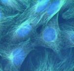
Microtubules are always present in a cell, acting as a cytoskeleton and providing structure. However, during mitosis this cytoskeletal array is disassembled and microtubules form running between two points at opposite sides of the cell called centrosomes. This structure is known as a bipolar spindle assembly.
The microtubules continually grow out and shrink back from the centrosomes, probing the cell until they collide with a chromatid and bind to it via an attachment point in the centre of the chromatid - a centromere. The microtubule then transports the chromosome released from the chromatid to the centrosome before the cell divides.
For microtubules to grow and shrink rapidly while probing for chromatids, they must be intrinsically dynamic polymers capable of assembly and disassembly. Therefore, any agent that increases or decreases microtubule stability has a dramatic effect on mitosis.
All current treatments targeting microtubules to combat cancer work like this. At high concentration, vinca alkaloids such as colchicine and vinblastine, first introduced in the 1960s, destabilise microtubules and the bipolar spindle breaks down. For taxanes such as paclitaxel, more commonly known as taxol and introduced in 1986, the opposite is true; the microtubules are stabilised, preventing the cell from dismantling the spindle fibres - halting the mitotic cycle. Though the exact trigger is still unknown, when mitosis stops for long periods the cell releases a signal and apoptosis occurs; the cell destroys itself.
The effect of these drugs on microtubules is a potent weapon in the fight against cancers, but while the target is known, exactly how the chemicals cause stabilisation or destabilisation of microtubules is not. It is critical, for many reasons, to know this exact mode of action.
Natural product development
All treatments currently in use are based on natural products; the vinca alkaloids come from the periwinkle plant and taxol comes from the Pacific yew tree. Developing more potent synthetic analogues demands an insight into what part of these complex natural molecules causes their activity.
Current treatments have some undesirable side effects. These include: decreased blood cell production; higher risks of infection; changes in heart function; and reversible nerve damage. If treatments are understood better, side effects can be predicted, controlled and, hopefully, minimised.
But before the methods of action of these drugs are assessed an understanding of what causes microtubules to be dynamic in the absence of drug molecules is needed.
Marcel Knossow and his colleagues at the Laboratoire d’Enzymologie et Biologie Structurale, Gif sur Yvette, France, are one group of researchers studying microtubule dynamics.
’Tubulin is unusual in the way it stochastically alternates phases of growth and disassembly to form microtubules, which is an important feature of the dynamic architecture of cells,’ Knossow explains. ’Our goal is to understand how microtubule dynamics are regulated from a structural point of view.’
Microtubules and monomers
Microtubules are made up of monomers called tubulin - heterodimers composed of tightly bound α-tubulin and β-tubulin monomers and two bound molecules of guanosine triphosphate (GTP). The α- and β-tubulin are arranged with one molecule of GTP trapped between the two tubulin subunits, and the second located on the surface of the β-tubulin molecule.
These make head-to-tail polymers, forming a protofilament: a long string of tubulin heterodimers. These in turn line up side by side, with β-tubulin molecules in line with other β-tubulin units. In vivo, 13 protofilaments generally congregate to form the hollow, rod-shaped microtubule.
As a consequence of this head-to-tail association, one end of the microtubule consists solely of α-tubulin, while the other consists only of β-tubulin. The β-tubulin end, or ’plus’ end, is more dynamic than the α-tubulin ’minus’ end. Microtubules grow and shrink more rapidly at the plus end - the minus end is normally attached to the centrosome, so long as the tubulin concentration stays above a critical level.
But even above this critical concentration, microtubule ends can suddenly stop growing and begin to shrink rapidly - a process aptly called a ’catastrophe’. Stops in growth or shrinkage are called ’pauses’, and regrowth is known as a ’rescue’.
It might be the GTP molecules that make the plus end more dynamic. GTP is only available at the plus end on β-tubulin, because the second GTP molecule in the heterodimer is buried between the α-tubulin and β-tubulin subunits. When the heterodimers polymerise, GTP quickly hydrolyses to guanosine diphosphate (GDP), which keeps the accessible GTP at the plus end only.
"Microtubule ends can suddenly stop growing and begin to shrink rapidly - a catastrophe"
This hydrolysis is also thought to cause the microtubules’ dynamic instability; with GTP hydrolysis, a length reduction of two to four per cent is seen in the tubulin heterodimer. This is indicative of conformational changes that induce strain in the protofilaments and make microtubules unstable.
This research shows that drug molecules interfering with GTP hydrolysis will reduce the dynamics of microtubule assembly. It also shows that current anti-cancer agents are active either by destabilising interactions between heterodimers in protofilaments, or by changing the conformation of strain-removing protofilaments and stabilising the polymer.
But to fully understand the action of anti-cancer drugs in use now, and to design new drugs with similar or better activity, the exact binding sites occupied by molecules on or within the tubulin heterodimer have to be identified.
To do this, tubulin’s exact structure was needed. This was achieved only after 25 years work - the structure was finally published in 1998, and a refined version was published in 2001.
Structure solving for binding sites
Conversely, taxol’s structure has been solved many times by x-ray crystallography and NMR analysis. Comparing these two structures, to work out taxol’s binding site, showed the taxol molecule occupying a pocket in β-tubulin that stabilises microtubules and therefore inhibits chromosome transport in dividing cells.
But the presence of this binding site raised an interesting question: since taxol and similar compounds are not found naturally in cells, why does a binding pocket on β-tubulin exist? Linda Amos and her colleagues at the MRC laboratory of molecular biology in Cambridge, UK, have been studying tubulin’s atomic structure and examining natural cell contents to suggest what the binding site might naturally be for.
"Microtubule-stabilising drugs were taking the place of natural stabilising agents found in most cells"
’The binding site is very highly conserved,’ says Amos, ’it seemed likely therefore that the microtubule-stabilising drugs were taking the place of some natural stabilising agents found in most cells.’
Amos determined that one possible candidate is microtubule-associated proteins (MAPs). One widespread group of these MAPs, which includes mammalian proteins MAP2 and tau, bind to a site on β-tubulin that overlaps with taxol’s binding site.
Stabilising microtubules
More evidence for this similar binding position came from competition studies: the amount of tau bound to microtubules was less in the presence of taxol and discodermalide than in their absence.
Amos suggests that the MAPs’ role is important in not only stabilising microtubules, but also in determining the spacing between microtubules and providing additional binding sites.
When Amos compared the structure of the tubulin heterodimer from mammals with FtsZ, a monomeric homologue of tubulin from the bacterium Methanococcus jannaschii, she found a similar binding pocket in the same position as the pocket in mammalian tubulin. Since anti-cancer drugs bind to this pocket in tubulin and cause cell death, could a new type of antibiotic drug be found that binds to FtsZ and also causes apoptosis?
Amos thinks it is possible: ’A compound that was small enough to pass through the cell membrane and that would bind tightly and specifically to FtsZ and not to tubulin might be an excellent antibiotic. However, at present, no candidate compounds are known.’
Future drug design
So how will this detailed knowledge of tubulin and microtubules affect the future of anti-cancer drug design?
Research carried out so far shows that all current drug molecules occupy similar binding sites on the tubulin heterodimer. Knowing this should help in designing new compounds with three-dimensional structures similar to current drugs, which will be active in the fight against cancer.
’The current structural data will help pharmaceutical chemists design new drugs that preserve tubulin-binding activity but lack the common problems with drugs of this class,’ says Knossow.
Only more research will lead to accurate predictions of the activity and potency of new drugs. ’More knowledge about the structure of complexes formed between tubulin and anti-cancer drugs will greatly help for novel drug design,’ Knossow explains. Amos adds: ’It seems likely that the shape of the binding pocket varies slightly when different drugs are bound. Molecular modellers would need to learn what sort of interactions are possible before accurate predictions about new compounds could be made.’
Before this is possible, higher resolution data for tubulin’s structure is needed. Mammalian brain tubulin is used at the moment, because it is easily purified in an active state. But it is a mixture of isoforms and is subjected to post-translational modifications.
Alternative tubulins are under investigation and, while work remains to be done, it is evident that better understanding about the way molecules control microtubule dynamics means better design of drugs and better treatments in the ongoing battle with cancer.
Andrew West is a science writer and post-doctoral researcher at Queen’s University Belfast, UK


No comments yet