Nuclear magnetic resonance spectroscopy is solving the 3D structure of previously inaccessible protein structures, thanks to recent advances in the field. David Bradley reports.
For decades x-rays dominated protein characterisation. All the big names fell for its crystallographic charms - myoglobin and haemoglobin, insulin, DNA and RNA, and dozens of enzymes. But, as the protein databanks filled, researchers realised that many protein crystal structures were seemingly inaccessible to them for one simple reason - they couldn’t be crystallised.
With no crystal, crystallography fell from grace. New character witnesses would have to be called on to offer evidence of protein power. Techniques such as nuclear magnetic resonance (NMR), infrared and ultraviolet/visible spectroscopy and mass spectrometry, once game only for hooking small molecules, entered the protein characterisation field.
Why is there such a great desire to know a protein’s detailed structure? Proteins, whether enzymes, receptors, or transport proteins, underlie almost every facet of life. When they are blocked or over-stimulated, or when a pathogen’s proteins break through our immune defences, the result is disease. A clearer picture of the proteins involved can provide a therapeutic target. Finding the means to unblock an inhibited protein, inhibit an overactive one, or eliminate otherwise dysfunctional proteins is key to treating countless diseases, from bird flu to the fungal skin disease zygomycosis.
Over the years, crystallography has provided biomedical researchers with the requisite atom-by-atom structures of countless protein targets. However, the structures of a large number of proteins, many of which do not crystallise, have yet to be solved.
Spectroscopic methods, which work on molecules in solution as well as in the solid state, are coming to the fore to help researchers unravel protein folds.
Even these have drawbacks. Mass spectrometry, for example, can reveal which protein is present in a sample and how much of it there is, but cannot tell us anything about what the protein looks like in its folded or active state: its tertiary structure.
Protein structures that resonate
In the mid-1970s, Kurt Wüthrich from the Swiss federal institute of technology (ETH) in Zurich, began to think about how NMR, which probes atomic spin using a static magnetic field and an applied radio frequency, might provide new insights into tertiary protein structure.
This analytical technique, well known to chemists who use it to reveal the structure of small molecules in solution, seemed perfectly suited to studying proteins not amenable to x-ray techniques. Most proteins of interest from a biomedical research perspective exist in solution in some form or another rather than as isolated crystals. After all, it is the proteins found in bodily fluids and those that nestle in the myriad cell membranes that are of most interest.
Moreover, NMR has another trump card to play because sophisticated upgrades to the basic spectrum can also reveal how a protein changes with time or in response to the presence of other chemicals; it can reveal a protein’s dynamic characteristics and conformational equilibria.
Changes such as protein folding, aromatic ring flips, amide proton exchange, hydration and protein interactions with DNA can all now be unveiled using protein NMR.
Wüthrich was awarded the 2002 Nobel prize for chemistry for using NMR to determine the 3D structure of biological macromolecules in solution. His approach, a framework for which was formulated in 1982, was based on sequentially assigning each hydrogen signal in a protein’s NMR spectrum. This provides the cornerstone of protein NMR, allowing distances between pairs of the scores of hydrogen atoms in almost every protein to be calculated and so reveal the 3D structures.
The technique works for protein solutions and even proteins in living cells and in 1985 was used to obtain the complete structure of a globular protein, bovine seminal proteinase inhibitor.
Wüthrich’s research group has solved some 50 protein structures this way, including protein complexes of tendamistat, an amylase inhibitor, metallothionein, a protein that transports metals, and prion proteins.
Protein NMR has moved on apace since those early days and current estimates suggest that 15-20 per cent of all the thousands of known protein structures have been determined by NMR.
Where’s the motif?
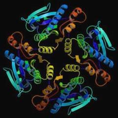
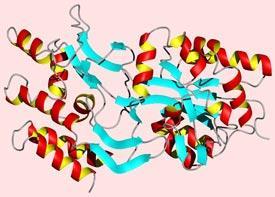
Although nature can create almost unlimited variations on protein sequences, a limited number of ’motifs’ appear in folded proteins. For example, the helix-turn-helix motif comprises two coiled sequences of amino acids, α-helices, joined through a short sequence that points the helices in the opposite direction to each other. Other motifs include the β ribbon, the zinc finger, and the Greek key.
In addition to these common structural motifs there are striking sub-structures, known as domains or modules, which biology uses repeatedly to perform many different functions. For example modules containing Greek keys, which allow two or more β-pleated sheets to sandwich together, are used in a wide range of proteins including antibodies in the immune system. Because these modules are relatively small (fewer than 300 amino acids) NMR is useful for determining their 3D structure.
The technique provides complementary information to x-rays and is useful for revealing binding interactions and dynamic information that is not available from diffraction techniques alone.
Automatic for the people
In March 2005 an international team of researchers took the first steps towards automating peak assignment, which could accelerate considerably NMR’s application to protein structure determination.
Peak assignment boils down to associating a group of spin systems in the NMR spectrum - ie a group of hydrogen atoms that are close together although not necessarily connected by neighbouring carbon, nitrogen or oxygen atoms - with the protein sequence of amino acids to indicate how the amino acids at different points along a protein chain fold together. Automating this process is a serious challenge in protein NMR.
Guohui Lin of the University of Alberta, Canada, and colleagues in Italy, Japan, and the US, have developed an algorithm that can estimate the proximity of specific amino acids in the protein chain based on the spin systems. They combined this algorithm with a fast computational filtering strategy, which provides optimal peak assignments automatically, potentially saving days or weeks of work.
They have tested the algorithm on a target protein from the Research Collaboratory for Structural Bioinformatics protein data bank and found that they can accurately and rapidly identify protein folds. The researchers expect that such a procedure will prove to be ’highly effective for fast and accurate protein fold and backbone structure predictions, using NMR data from only a small number of NMR experiments’.
Lin told Chemistry World that the team has tested the algorithms on almost 500 proteins using the NMR data in BioMagResBank. ’The average running time of our most recent integer programming based assignment algorithm is less than two seconds on a normal desktop computer,’ says Lin. ’The average assignment accuracy is about 94 per cent.’
Lin adds that ’unlike several in-house tools, our assignment algorithms return solutions within seconds and thus confirm that NMR protein structure determination can be automated and become high-throughput’. Moreover, the assignment algorithms are optimal with respect to NMR spectral data quality.
The team hopes to make the algorithms more fault tolerant by providing statistical evaluation on individual mappings, and to incorporate the assignment algorithms in the spectral peak picking to filter out fake resonance peaks.
Noesy adds up
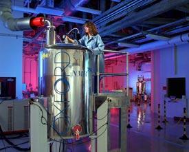
Industry is continually improving NMR spectrometers, developing higher field strengths, and bigger and more powerful superconducting magnets to squeeze every ounce of sensitivity from a spectrum.
Developments at the spectral interpretation stage continue to offer new clarity in protein structure determination.
Researchers at Carnegie Mellon University in Pittsburgh, US, have reported a different type of algorithm that can analyse the unassigned peaks in an NMR spectrum resulting from nuclear Overhauser effect spectroscopy (Noesy) and then piece together a protein structure from the fragments.
Noesy spectra are two-dimensional and reveal chemical details that conventional one-dimensional spectra fail to pick up.
Kurt Wüthrich and his ETH colleague Richard Ernst, who was awarded the 1991 Nobel prize for chemistry for his work on high resolution NMR spectroscopy, pioneered the use of Noesy for studying biological macromolecules in the mid-1970s.
The technique is not perfect. According to Miguel Llinás from Carnegie Mellon, the ’lingering problem’ with determining protein structures using NMR is the degree of subjectivity inherent in interpreting the spectrum.
In an effort to reduce this subjectivity, Llinás and colleagues are developing a robust algorithm to allow them to interpret spectra quickly using minimal data.
One such algorithm called Bacus uses a Bayesian statistical approach to analyse routinely nuclear Overhauser cross-peaks so that sequences can be assigned.
When testing a specific Bacus protocol for analysing NMR data, Llinás and colleagues reasoned that considering molecular ’fragments’ as building blocks rather than looking at each atom in isolation might allow them to speed up the analysis.
The distances between these fragments as revealed by Noesy could then be used to produce a likely bundle of protein structures through applied Bacus (Abacus).
The researchers carried out a blind test on NMR data from a 70-amino acid protein called mth1743 from the microbe Methanobacterium thermoautotrophicum. This revealed the Abacus protocol to be ’robust and rapid, and flexible enough to be readily adapted to other types of experimental heteronuclear-edited NMR data’.
This test protein is of interest to structural genomic scientists, but Llinás and colleagues generally focus on proteins such as those involved in blood coagulation, cell matrix metalloproteinases, and proteins concerned with neurological function.
Sail away
Masatsune Kainosho from Tokyo Metropolitan University, Japan, and colleagues are using isotopic labels to reduce line broadening in their protein NMR spectra.
Using stereo-array isotope labelling (Sail) they embed a complete stereospecific and regiospecific pattern of stable isotopic labels into a protein.
They recently demonstrated - for the 17kDa protein calmodulin and a 41kDa maltodextrin-binding protein - that Sail not only sharpens spectral lines but also simplifies spectra.
This allows them to solve high-quality solution structures for proteins twice as large as those commonly accessible using NMR. ’It thus makes a large class of proteins newly accessible to detailed solution structure determination,’
says Kainosho.
He told Chemistry World that the team is currently working on further optimised Sail patterns, which could enable NMR to determine high-quality solution structures of proteins up to 100 kDa and membrane protein structures.
’The impact of Sail on biomedical science results from the fact that among these proteins there are various disease-related potential drug targets that have been difficult to study up to now because they do not crystallise and are too large for classic NMR structure analysis,’ says Kainosho.
A solid approach
There remains no reason why NMR spectroscopists cannot complement x-ray crystallography data with solid-state NMR results. The techniques could provide unique insights into insoluble or non-crystalline biomolecules at the atomic level.
Marc Baldus and colleagues at the Max Planck institute for biophysical chemistry in Göttingen, Germany, explain how solid-state NMR offers protein scientists high-resolution conditions. These are obtained by spinning the sample in an ultra-high magnetic field close to 20 Tesla and using tailored pulses of radio frequency radiation to extract the key spectroscopic data. The angle at which the sample is spun, the magic angle, reduces the spectral line width by averaging out the magnetic field across the whole sample.
By refining their use of Mas (magic-angle spinning) NMR through the use of multiply or uniformly [13 C,15 N]-labelled proteins, for instance, researchers have been able to reveal the finer details of more complex structures.
Mas NMR comes into its own for studying protein aggregates. Proteins that clump together form an altogether different target for medicine than the usual active and dysfunctional proteins that have either to be blocked or triggered.
Clumps of proteins and misfolded proteins underlie diseases such as Alzheimer’s disease where proteinaceous plaques damage brain cells, lupus in which faulty proteins accumulate in cells to cause organ damage, and the spongiform encephalopathies (BSE, scrapie, Creutzfeldt-Jakob disease, etc) where misfolded proteins self-replicate and accumulate.
Mas NMR can also focus sharply on proteins bound to cell membranes, an incredibly diverse and important group of proteins involved in transporting chemicals in and out of the cell. Allowing researchers to study such proteins still bound to a membrane, essentially in the solid state, and more critically carrying a ligand, may reveal new insights into the dynamics of transport and signalling processes.
Even the most ardent NMR fan will admit that the technique can currently take protein characterisation only so far. Nevertheless, it has the significant advantage of revealing information about proteins in their natural state - whether in solution or membrane bound - and does not require them to be crystallised.
NMR complements x-ray studies perfectly and extends the range significantly. It would seem that science has seen through the dominance of x-rays and emerged with a powerful new set of tools.
David Bradley is a science writer
Companies
- Bruker Biospin, based in Massachusetts, US, sells a range of NMR equipment, including spectrometers. The company provides superconducting NMR magnets up to 900MHz. It also sells a range of NMR accessories, including cryoprobes and magic angle spinning probes.
- Oxford Instruments is based in Oxfordshire, UK. The company sells NMR magnets for ultra-high field applications, offering NMR magnetic field strengths up to 950MHz. It also sells benchtop NMR machines and accessories.
- Varian sells NMR magnets and a range of probes including cryoprobes and solid and liquid state probes. It also sells software to deal with data acquisition, processing and analysis.The company is based in Palo Alto, US.
- JEOL, based in Tokyo, Japan, provides a range of NMR spectrometers. Its ECX range is available from 300 to 500 MHz field strengths. Meanwhile, its ECA NMR covers 300 to 900MHz. The company also sells NMR probes.
- Goss Scientific Instruments is based in Essex, UK, and sells a range of NMR-related products including NMR probes, solvents, stable isotopes and sample tubes.
- NMRtec is a spin-off from Montpellier University, France. It offers contract NMR analytical services and has the facilities for solid and liquid state NMR. It also has a range of software packages.
- Cortecnet is a French web portal for NMR products, including deuterated solvents, NMR tubes and accessories.
- Intertek Caleb Brett is part of Canadian firm Intertek. The company carries out a range of NMR analyses and runs a UK service from Manchester.
- Mestrelab is a Spanish firm that provides NMR software including data prediction and analysis packages. The company also stocks virtual NMR simulator packages for students. Most of the software can be downloaded from its website.
Additional information
- Z-Z Chen et al, J. Comput. Biol., 2005, 12, 129
- M Kainosho et al, Nature, 2006, 440, 52
- S Luca et al, Acc. Chem. Res., 2003, 36, 858


No comments yet