Raman spectroscopy detects tumours after 'staining' with nanoparticles
A new imaging technique based on Raman spectroscopy has been used to illuminate tumours in mice with unprecedented precision [1]. If the technique can be safely extended to humans it could be a fast and accurate way to spot cancerous cells.
Whereas existing methods such as MRI and PET-scanning rely on large magnets or radioactive isotopes, Raman spectroscopy only needs a modified laser microscope and an injection of nanoparticles to act as a ’beacon’.
The Raman effect occurs when laser light is scattered by molecules - producing a unique spectral fingerprint for different compounds. But this signal is typically weak and has so far prevented Raman spectroscopy from being used to scan deep within the body.
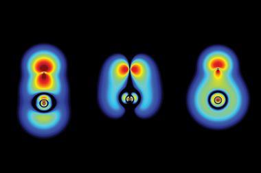
To solve this problem the team led by Sanjiv Gambhir at the Stanford University School of Medicine, California, made use of Surface Enhanced Raman Scattering (SERS): a process that uses selected molecules to boost signal strength.
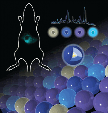
Two different boosters were used - gold-based nanoparticles and carbon nanotubes - which were attached to a tumour-seeking peptide and tracked as they moved around the body. The team was able to image nanoparticles in mice at concentrations of close to one-trillionth of a mole - 1000 times better than existing techniques.
The Raman signal also reveals more than just the location of the particles - it is able to distinguish between nanoparticles with slightly-differing structures, which could be a useful tool for future toxicity studies, for example.
Critical to the study was a modified Raman laser microscope designed to detect the Raman nanoparticles. The team believes it should also be possible to use other Raman-active nanoparticles and tumour-seeking systems such as monoclonal antibodies with their system and are now working to optimise particle sizes and dosages.
A clinical trial has already been planned to search for early-stage colorectal cancer using this technique. In a second study also published this week, Gambhir and colleagues have shown that carbon nanotubes appear to be non-toxic in mice [2], which is good news for the Raman team.
’This is a very nice demonstration of deep tissue imaging,’ says Ioan Notingher, an expert in analysing biological molecules with Raman spectroscopy at the University of Nottingham. ’The depth is suitable for many applications and detection limits are very good, but the toxicity of the nanoparticles needs to be carefully evaluated to make this truly non-invasive.’
Lewis Brindley
References
et alProc. Natl. Acad. Sci.et alNat. Nanotech., 2008, DOI:10.1038/nnano.2008.68


No comments yet