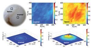
Dental cavities come about when lactic acid, produced by oral bacterial, decomposes the protective mineral coating on teeth, leaving them vulnerable to decay. Detecting signs of rotting early is vital for successful treatment to prevent painful infections and loss of teeth, but they are often hard for a dentist to spot through visual inspection or x-ray images.
Raman spectroscopy is commonly used to assess the mineral content of bodily tissues as it is very sensitive to mineral crystals. However, classical Raman imaging uses linear laser light which produces an array of spectra and requires the sample to be moved with respect to the laser. This is time consuming and limits its clinically feasible.
Wide-field Raman imaging is being investigated by Ozan Akkus, and his team at Case Western Reserve University, as a more efficient method. Their most recent strategy combines wide-field Raman imaging with near infra-red lasers, special filters and 2D-charge-coupled device (CCD) cameras. It differs from classical Raman imaging as it only produces images of select wavenumbers of interest and defocusses the laser beam so larger areas can be analysed.
Areas of low and high mineral content are identified as weak and intense Raman signals, respectively, in a process that is 13 times faster than previously used Raman methods. Akkus stresses that ‘if dentists were able to image the mineral variation over the entire surface of a tooth, they would be able to identify and address any emerging problems’.
Jemma Kerns, a Raman spectroscopy expert at University College London, UK, comments that this application of Raman spectroscopic imaging, to identify mineralisation changes in dental lesions, is significant and will ‘allow for the development of better treatment options to directly benefit patients in the future.’
Akkus says that they now wish to increase the area that they can image and reduce the cost of the procedure in order to translate this concept into practice.
References
This paper is free to access until 22 May 2014. Download it here:


No comments yet