Transmission electron microscope image shows direct visualisation of DNA bundle
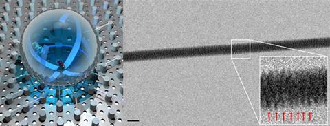
To suspend the fibres, the team exploited the tendency for surfaces covered with regular arrays of microscopic features to be superhydrophobic. This allowed them to place droplets of aqueous DNA fibre solutions on the surface. As the droplets dry, the DNA is left behind, stretched between the pillars.
The silicon substrate below has a hole in between each pair of pillars, through which the transmission electron microscope electron beam could be focused to image the fibres. This allowed the team to clearly visualise and measure the periodic features of the DNA strands that make up the fibres, particularly on the strand closest to the edge of the bundle.


No comments yet