Tropical frogs create remarkable foams to protect their spawn. Exploration of the underlying chemistry has only just begun, as Michael Gross discovers
When physical chemist Alan Cooper goes looking for samples to analyse in his laboratory, he may tiptoe around an old graveyard in Trinidad at night, careful not to disturb the sleeping water buffalo employed to keep the grass short. The buffalo’s wallow attracts túngara frogs (Engystomops pustulosus), which like to deposit their spawn there.
What attracts Cooper and his colleagues, however, is the intriguing way in which these frogs spawn. ’The female produces clutches of eggs together with a foam precursor liquid. The male, clinging to her back and using his legs in a rapid "egg-beater" motion, whips the liquid into a foamy mass while fertilising the eggs,’ Cooper explains. The resulting bug-infested foam nest has roughly the consistency of beaten egg white. Amazingly, however, it can last for several days even in tropical heat and rain. If researchers remove the eggs, the foam remains stable for more than a week.
Cooper and co-workers at the University of Glasgow, UK, are studying frog foam nests to find out which biomolecules provide it with its remarkable stability and spawn-protecting ability. Taking care not to deprive any unborn frogs of a happy life in the mud, they collect foam nests from sites where the spawn would have been doomed in any case - as in the graveyard, where it would most likely be flattened by the water buffalo the next day.
Cooper started this investigation in 1997, together with biologist Malcolm Kennedy. At the time, the Wellcome Trust launched an initiative to fund unusual projects that might otherwise not get supported, and the Glasgow-based team was successful with their grant application. Subsequent funding for the project was intermittent, says Cooper, which, together with the scarcity of raw material, is why it has taken a number of years to come to fruition.
A family of foam proteins
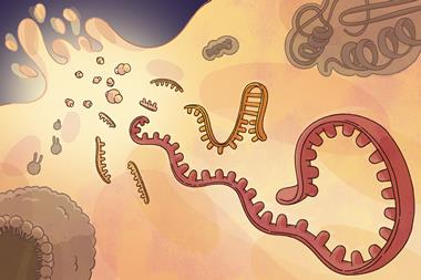
But it was worth the wait, says Cooper, as he and his colleagues have since revealed a whole new family of proteins responsible for the unusual properties of frog foam, which they have named ranaspumins, derived from the latin words for ’frog’ (rana) and ’foam’ (spuma).1, 2 Physico-chemical investigations showed that these proteins accumulate at the air-water interface, thus stabilising the foam.1 This is very surprising behaviour - one of the first things anybody handling proteins in the laboratory learns is that foaming is bad news. The foaming usually indicates that the protein has lost its native structure, and rearranged such that its hydrophobic parts - normally tucked away in the inner core of the structure - have reoriented themselves towards the air bubble, while the hydrophilic ones remain anchored in the water phase. If a laboratory sample starts foaming, its owner will probably find that whatever protein activity existed before will have disappeared.
Given the frog proteins’ unusual behaviour, the researchers set out to study them in more detail, to work out what was behind their remarkable stability and surfactant properties. They found that the foaming liquid is a dilute (1-2 mg ml-1) solution of proteins and complex carbohydrates. While the carbohydrates are still uncharacterised, the researchers managed to identify six different proteins in the foam nests of E. pustulosus - ranaspumins (Rsn) one to six.
Four of the proteins, Rsn-3 to Rsn-6, turned out to be members of the lectin family, a large group of carbohydrate-recognising proteins, first discovered in plants but now known in all kinds of organisms. Tests on these ranaspumins revealed that they do indeed bind carbohydrates - Cooper and his team suspect that they hook up the surfactant proteins to a stabilising network of carbohydrates present in the foam, as well as inhibiting microbial growth and deterring predators. ’Lectins can agglutinate cells - that is, make them stick together - and this can inhibit microbial proliferation,’ Cooper explains. ’Lectins can also act as anti-feedants and give predators a bellyache, so they don’t come back again.’
Of the remaining two proteins, Rsn-1 appears to be related to the cystatins. This family usually inhibits protein-degrading protease enzymes - the researchers have been unable to detect such activity in the frog protein, although the natural foam cocktail does inhibit protease activity. Finally, Rsn-2 displayed an amino acid sequence unrelated to any other protein known to science, which is why Cooper and his colleagues scrutinised its structure more closely. Experiments showed that this protein is most effective in reducing surface tension and producing foam, even though in the absence of the other components, the foam doesn’t last very long.
The novel surfactant
Using NMR spectroscopy, collaborators at Glasgow - Brian Smith and Cameron Mackenzie - solved the structure of Rsn-2, and found that, like Rsn-1, it appeared to have a cystatin-like folding pattern.3 The structure, which is typical of cystatins, consists of a flat beta-pleated sheet with a single alpha helix packed on top of it.
Large parts of the protein chain at both ends appear to be unstructured in solution, which may be a clue to its surfactant properties. While it is known that completely unfolded (denatured) proteins tend to foam, the ranaspumins display surfactant properties at much lower concentrations than ordinary denatured proteins would, so there is still some explaining left to do.
A link between surfactant properties and unstructured regions wouldn’t be surprising, points out Véronique Receveur-Bréchot, who studies intrinsically unstructured proteins at the French National centre for research (CNRS) unit for NMR spectroscopy in Marseille. ’Beta casein in milk, for instance, was one of the first intrinsically disordered proteins to be discovered. By acting as a surfactant, it allows the formation of micelles in milk [which solubilise important nutrients such as calcium phosphate],’ she explains. ’Intrinsically unstructured proteins often have a high proportion of charged or polar amino acids. In combination with a hydrophobic domain, this can lead to amphiphilic, surface active properties.’
As yet, the exact conformation Rsn-2 adopts as it accumulates at the foam’s air-water interface is still to be determined. But based on its cystatin-like solution structure, Cooper suggests that the Rsn-2 molecule, on approaching the interface, opens up with a hinge-like movement, separating the helix from the sheet, and thus exposing hydrophobic parts of the molecule to the interface. ’The unstructured, but amphiphilic N- and C-terminal regions might provide the driving force, pulling the "hinge" open at the air-water interface,’ says Cooper.
In collaboration with colleagues in Halle, Germany, and Manchester, UK, Cooper has used surface infrared spectroscopy and neutron reflection experiments, which support this mechanism. To test it more directly, his group is now carrying out mutagenesis of Rsn-2, aiming to remove the unstructured regions, or to engineer in a disulfide crosslink to fix the hinge in a closed position.
Blue as a smurf can be
Apart from the túngara frogs, there are several other tropical frog species producing biofoams to protect their spawn. Among them, Polypedates leucomystax stands out because its foam nests turn blue-green after a while. Cooper and colleagues have studied these foam nests on field trips to Malaysia and have recently identified the cause of this colour as a highly unusual protein, which they named after the similarly coloured cartoon heroes: ranasmurfin.4
Ranasmurfin readily forms deep blue crystals of such good quality that the researchers, in collaboration with Jim Naismith and colleagues at the Scottish Structural Proteomics Facility at the University of St Andrews, UK, didn’t even need to solve its amino acid sequence to figure out what they were looking at. Unusually for a protein crystal structure, they could identify amino acids directly from the electron density map derived from the X-ray diffraction patterns. Any ambiguities were quickly resolved by mass spectrometry.
The researchers discovered the protein had a novel folding pattern, which is very rare these days, as there are more than 50,000 protein structures known, and experts believe that there are only a few thousand fundamentally different folds. They also discovered a novel cross-link between the two subunits, namely an indophenol-like group which has never before been observed in a stable protein structure.
Using spectroscopic studies of the protein and of model substances, Cooper’s team showed that this unusual group is also the source of the characteristic blue colour. It arises by post-translational modification - a chemical reaction between amino acid residues on the protein chain that occurs after the protein has been synthesised and folded into its native conformation, much like the chromophore in Green Fluorescent Protein (GFP), the widely used gene marker whose discovery was honoured with the 2008 Nobel prize in chemistry.
But while the chemistry behind the colour of the ranasmurfin protein appears to be clear, no obvious biological reason for its smurf-like shade has yet emerged to explain why it has evolved to be so colourful. Conceivably, the indophenol group could just be there to stabilise the protein in the unusually stressful environment of a foaming solution, and the colour might just be a side effect of the chemistry. Alternatively, it may have evolved to shelter the spawn from sunlight, or to confuse predators.
Biofoams everywhere
This pioneering interdisciplinary work has inspired other researchers to look more closely at the foam nests, which have so far only been known as curious by-products of frog reproduction. Vania Melo at the Federal University of Ceará at Fortaleza, Brazil, is now applying similar analyses to the foam nests of tropical frogs found in northeast Brazil, and has identified a surfactant from Leptodactylus vastus, Lv-ranaspumin.5’Lv-ranaspumin is a surfactant protein that comprises almost 90 per cent of the bulk protein of the foam,’ Melo explains. ’It shows no amino acid sequence similarity with the ranaspumins reported by Alan Cooper’s team, suggesting a great diversity of these ranaspumins. Foam nests appear to have evolved independently in different phylogenetic lineages.’
Working on the opposite side of the South American continent, on the Chilean coast, Juan Carlos Castilla and his team at Santiago de Chile have recently shown that foaming also protects the spawn of marine organisms, such as the intertidal tunicate Pyura praeputialis.6
’We have evidence that biofoams in the rocky shore are, if not common, at least produced by a number of invertebrate species and phyla,’ Castilla says. ’We have field evidence for biofoam production in Chile for one species of starfish and a chiton (mollusc). Biofoams in the rocky intertidal zone may be produced to retain eggs at early developmental stages, as in frogs.’ Unlike the frog foam nests, however, marine biofoams are thought to arise from carbohydrate surfactants - no protein surfactant has yet been identified in the marine context.
In other biological settings, such as the human lungs, lipids act as surfactants to produce biofoams. Horse sweat is an interesting non-amphibian biofoam that also uses proteins as surfactants. The Cooper-Kennedy team is studying these proteins, the latherins, and first results are due to appear soon.7
Useful foams
Considering the frog foam’s remarkable ability to protect sensitive eggs and embryonic stages in a harsh environment teeming with microbes, one would immediately think of potential medical applications, for instance in wound dressing, surgical fillings, or matrices for tissue regeneration.
Together with Helen Grant at the University of Strathclyde, UK, Cooper has begun to explore the application potential of biofoams. Preliminary experiments showed that the frog nest material acts as a kind of biological Teflon: cells simply don’t stick to the material.
Other applications can be imagined in food, laundry, and environmental decontamination technology. However, if uses never emerge, it will still be useful to have found out intriguing details about the reproduction of frogs. With the threat of extinctions caused by climate change, such knowledge can help to save species. Timothy Wess, a biophysicist at the University of Cardiff, UK, comments: ’I’ve followed the developments that they have made at the interface between reproductive zoology and protein chemistry, as it leads towards possible advances in biomedicine. Whatever happens next, it shows that we need to support curiosity driven research. This is a fascinating and entertaining journey, and if you can pack in a new protein fold, novel crosslink and chromophore on the way, so much the better.’
Michael Gross is a science writer based in Oxford, UK
References
1 A Cooper et al, Biophys. J., 2005, 88, 2114
2 R I Fleming et al, Proc. R. Soc. B, 2009, DOI: 10.1098/rspb.2008.1939
3 C Mackenzie et al, Biophys. J., 2009, in press
4 M Oke et al, Angew. Chem. Int. Ed., 2008, 47, 7853 (DOI: 10.1002/anie.200802901)
5 D C Hissa et al, J. Exp. Biol., 2008, 211, 2707
6 J C Castilla et al, Proc. Natl. Acad. Sci. USA, 2007, 104, 18120 (DOI: 10.1073/pnas.0708233104)
7 R E McDonald et al, PLoS ONE, 2009, in press


No comments yet