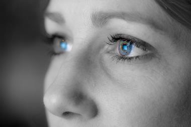
People can lose their eyesight for a number of different reasons but there are a few promising treatments on the horizon. Michael Gross looks them up.
People can lose their eyesight for a number of different reasons but there are a few promising treatments on the horizon. Michael Gross looks them up.
Globally, there are over 50 million blind people and, unless revolutionary cures are found, the number is predicted to rise by 50 per cent within the next two decades. Cataracts, a loss of vision resulting from protein aggregation in the eye lens, used to be one of the most common causes, especially in old age. The widespread use of artificial lens implants, first tested by Harold Ridley 55 years ago, has now brought this problem under control ( Chem. Br., January 2002, p30). Sight impairment resulting from eye disease outside the lens requires both chemical and biological approaches to therapy.

One of the most widespread causes of blindness is glaucoma. Essentially, it is a drainage problem affecting the liquid, known as the aqueous humour, contained in the front of the eye, between the cornea and the iris and lens. This liquid is constantly renewed and drained away through a network of holes called the trabecular meshwork. When drainage is blocked, the intraocular pressure within the eye increases, which deforms the surroundings of the optic nerve and thereby leads to loss of vision. Ophthalmologists often measure the intraocular pressure by directing a short puff of air into the eye and measuring the light reflected from the cornea. The brief deformation of the eye caused by the air jet allows the instrument to calculate the intraocular pressure. This method can pick up the first indications of glaucoma long before the patient notices any symptoms.
There are several treatment options for glaucoma, including a number of approved drugs that either reduce fluid production ( eg beta blockers such as timolol maleate) or improve drainage (typically prostaglandin analogues like latanoprost). Doctors can also improve drainage surgically by cutting new channels. In theory, this option can solve the problem in a one-off treatment, without the need for long-term medication. However, care needs to be taken to avoid the build-up of scar tissue that might hinder vision. Researchers at the Institute of Ophthalmology (IOO), at University College London, UK, are currently involved in clinical tests of several methods designed to prevent scarring, including the use of the drug 5-fluorouracil - a cytotoxic analogue of the RNA nucleobase uracil that is also used in cancer chemotherapy. The causes of glaucoma are believed to lie in a combination of genetic predisposition and certain environmental factors.
Accordingly, keeping a register of families carrying the gene is an important instrument to help identify sufferers early and - one day - treat them more efficiently. IOO researchers have studied RP over many years, mainly working with patients from the Moorfields Eye Hospital in London. This work has not only resulted in pinpointing several of the mutations that can trigger the disease, but also in the creation of a register holding the details of over 4000 affected families and over 30 000 individuals.
The disease involves a gradual loss of the photosensitive cells in the retina, normally progressing from the outside inwards. That is, patients first lose the highly sensitive (but colour blind) rod cells at the periphery, and may notice the loss of night vision. As the decay progresses towards the core of the retina (the macula), where the colour-sensing cones are more abundant, general vision deteriorates, and patients will typically be registered blind before they turn 40. At the moment, there is no approved treatment that can stop the progression of the disease towards total loss of vision.
However, a number of potential therapies are currently under development. Drugs known as survival factors, if regularly injected into the eye, could help prevent cell death in the retina and block the progress of the degeneration. These substances are typically regulatory proteins such as growth factors ( eg basic fibroblast growth factor, bFGF) and neurotrophic factors ( eg brain-derived neurotrophic factor, BDNF), which were found to protect photoreceptors from the damage induced by constant exposure to light.
As a disease with a clearly identified genetic cause, RP is an obvious candidate for gene therapy. However, delivery of ’replacement genes’ into human cells is still a challenge. Even the most successful gene therapy trial, which cured 10 so-called ’bubble babies’ of their severe immune deficiency, suffered a serious setback when two of the children developed leukaemia. Even though trials are to be continued on several severe diseases including RP, there is at the moment no way of knowing when these obstacles will be overcome.
Another silver lining for the patients affected by RP and even those who may have already lost their eyesight is the possibility of implanting replacement cells. While the availability of donor organs would never be sufficient to keep pace with such a relatively common disease, it may one day be possible to grow healthy retinal cells in a culture dish and implant them into the patient’s eye. Such experiments are currently being carried out for other vision-related type cells.
But then again, does one need a biological retina to convert light into neurochemical signals? The first trials of very primitive electronic retina analogues, containing as few as five by five pixels, have demonstrated that it is possible, in principle, for a man-made electronic device to feed signals to the brain that are processed as vision.
In a photochemical variation on this theme, Robert Givens’s group at the University of Kansas, US, has begun developing a retina prosthesis based on a chemically caged version of the neurotransmitter glutamate. When hit by light, the molecule liberates glutamate, which is essentially what healthy rods and cones do. Supplying the eye with a constant flow of the caged chemical will be the real challenge in the development of this ’chemical retina’.
While the precise mechanisms causing the characteristic symptoms of the disease are not yet fully understood, it is clear that the cause does not lie in the retinal photosensors per se, but rather in the adjacent layer, the retinal pigment epithelium (RPE). This layer is responsible for feeding the retina, and also for taking care of its waste, eg molecular compounds damaged by unwanted photo-oxidation reactions. In AMD patients, the RPE fails to keep up with the housekeeping work. As a consequence, waste accumulates between the layers, and cells begin to die. Further vision loss results from scarring processes involving the formation of unwanted blood vessels.
While there are genetic elements that may predispose people to AMD, a number of environmental factors including diet, smoking, and exposure to UV light are also under suspicion as contibutors to the condition. As for the treatment, there is currently no way of stopping the progress of the disease, but medics can try to block the unwanted scarring and formation of new blood vessels using laser or radiation treatment. Several drugs tested for the same purpose have failed due to their side effects. Research is being conducted with the long-term goal of implanting cells to regrow the RPE. These could come from organ donors, from RPE-cells held in culture, or indeed from nerves in other parts of the body.
Vision can also be affected by problems arising from the immune system, including allergies and autoimmune disease. A fairly common disease involving an autoimmune response against the retina is known as uveitis. It appears that the retinal blood vessels recruit white blood cells which then produce cytokines and other substances that can damage the retina. Further details and possible means of stopping the disease have yet to be uncovered.
Much like the over-enthusiastic immune system in uveitis, an under-achieving immune system weakened by Aids can spell trouble for the vision process. Infection of the retina with cytomegalovirus (CMV) is believed to affect around 30 per cent of HIV positive people at some stage of their lives. Research from the group of John Kempen at Johns Hopkins University, US, has recently demonstrated that a multi-drug therapy known as ’highly active antiretroviral therapy’ (HAART) not only saves the lives of Aids sufferers but also improves their eyesight, because it drastically reduces the incidence of CMV infection.
Acknowledgements
Michael Gross is science writer in residence at the school of crystallography, Birkbeck College, University of London. His latest book, Light and life is available from Oxford University Press.
Further Reading
- Oak Ridge Associated Universities (ORAU) website
- J H Kempen et al, Arch. Ophthalmol., 2003, 121, 466.
- N S Dejneka and J Bennett, Bioessays, 2001, 23, 662.
- Birkbeck University of London: EyeSite
A site for sore eyes
Researchers at two UK universities have set up a database that puts proteins of the eye into their evolutionary context and will be useful both for clinical and for fundamental research. EyeSite you with a larger-than-life ’interactive eye’, but behind that playfulness lies a serious scientific tool that half a dozen researchers have put together in several years.
Christine Slingsby, a specialist on eye lens proteins and principal investigator on the EyeSite project, recalls how the project evolved from a network of contacts rooted in the crystallography department at Birkbeck College, London. ’It all goes back to the times when Tom Blundell and Janet Thornton were here, doing structural biology and bioinformatics,’ she says. ’Then, when Janet moved to UCL [University College London], crystallographers over here stayed in touch with bioinformaticians at UCL’s biochemistry department, and thought about common projects.’ The Bloomsbury Centre for Structural Biology was one way in which contacts were strengthened. Then, five years ago, both departments obtained a joint grant from the Medical Research Council to develop databases including EyeSite.
Several PhD students and post-doctoral fellows helped develop the data handling programs and then collected the input data from existing public databases. They included not only every protein that is expressed in human or other vertebrate eyes, but also all other proteins that can be shown to have a significant degree of relatedness (homology) to eye proteins. Thus they ended up with over 70 000 sequences, grouped by EyeSite into 722 families. If detailed structures are known, the program will show them. If there isn’t a structure but a high degree of homology to a protein of known structure, the program can create a model (ie a reasonable guess) of the new protein based on the known structure.
How do users find their way in this enormous wealth of data? One can, of course, browse the database, asking it to show lists of proteins produced, for example, in the eye lens of humans, or in a rodent retina. The interactive eye on the front page also leads to such lists, which are created on demand, like in a search engine. More importantly, however, researchers with a novel protein or gene sequence, who suspect that it might have something to do with the eye, can use their raw data to search for any related sequences in the database. As the database contains a large fraction of all known sequences, they are likely to find something interesting even if their protein is not directly related to eye functions.
After a few years of using and improving a trial version of EyeSite the researchers have recently gone public by presenting the site in a special issue of Nucleic Acids Research on databases. So now the EyeSite can reach eyeballs all over the world.


No comments yet