New technique can view blood vessels' structure and molecular signs of ill health at the same time
An international team of medical researchers has developed a way to perform a check up on an artery’s physical structure and molecular markers of ill health by coupling a catheter with imaging technology. This device could help develop new drugs to stabilise arterial plaques and may one day assess the health and treatment options of patients with hardening arteries.
Fat, cholesterol, calcium and other substances in our blood form a plaque which builds up inside our arteries, restricting the flow of blood. Worse, plaques can rupture, and blood reacts to their contents by clotting - potentially causing a heart attack or stroke. ’It’s a healing response gone awry,’ says Farouc Jaffer, co-leader of the team with Gary Tearney, both at Massachusetts General Hospital and Harvard Medical School.
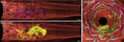
To investigate the health of arteries, doctors thread them with catheters containing imaging devices, but they have to choose between looking at the morphology of the artery, plaques and stents (implants which prop arteries open), or molecular signals of ill health. To see the structures and proteins in one go, the researchers probed arteries using a single optical fibre, connected to an infrared laser. The light through the central fibre builds a structural picture of the blood vessel from the way light scatters off the artery wall, while the secondary fibre surrounding the central one picks up fluorescent signals that are indicators of inflammation and cell growth.
The team used this device to image plaques and a stent in live rabbits. To investigate plaques, they injected the rabbits with a fluorochrome that is activated by cysteine proteases, enzymes which signal inflammation. In the images produced, ’the inflammatory protease activity can be mapped to the plaque,’ says Tearney.
With the ability to see both structure and activity, Jaffer says that researchers could understand plaques better and determine what puts them at risk of breaking open. ’Until we actually understand which plaques will rupture we can’t even test the right preventative therapies,’ he says.
The team also installed a stent in an artery that fed the leg of a rabbit. After injecting the rabbit with a cyanine fluorochrome that binds to fibrin, a protein involved in blood clotting, the team imaged the stent inside the artery. They could not only see the structure of the stent and the clots around it, but they could identify fibrin on the stent’s metal struts. The researchers propose that detailed knowledge about stents in human patients could help doctors decide whether anti-clotting medications are necessary to prevent arteries from healing themselves closed.
Zahi Fayad of the Mount Sinai School of Medicine, US, calls the device ’a very important advancement into the field of intra-arterial imaging’. He suggests that it could first serve as a tool to investigate whether novel drugs which alter plaque composition are effective in making plaques more stable - preventing catastrophic ruptures. Still, he warns that moving the technology from the lab to the clinic could be tricky.
Tearney is more optimistic. Because the device is sheathed in a catheter, which is identical to those used in the clinic, he predicts that it could be used to image patients’ arteries within one or two years.
Kate McAlpine
Interesting? Spread the word using the ’tools’ menu on the left
References
H Yoo et al, Nat. Med., 2011, DOI:10.1038/nm.2555


No comments yet