


Japanese scientists have shown that coating insect larvae with Tween-20, a common detergent, lets them survive the powerful vacuum inside an electron microscope. The technique could pave the way for high resolution live imaging.
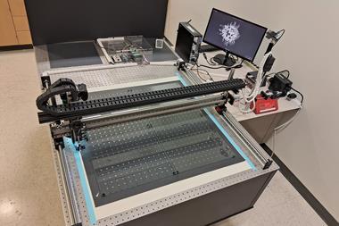
A grisly end awaits most living animals subjected to the high vacuum inside an electron microscope. The low pressure causes body water to evaporate, and death follows within minutes. But Takahiko Hariyama, of Hamamatsu University, and colleagues were surprised to find that fruit fly larvae remained alive and well in a scanning electron microscope (SEM) for over an hour.
High resolution imaging revealed a thin membrane covering each larva, suggesting that the bugs’ extra cellular substance (ECS) somehow protected them from the low pressure. However, when the same larvae were placed in a vacuum chamber without the electron beam from the SEM, they died.
‘ECS itself cannot protect the animal in a high vacuum,’ explains Hariyama, ‘but energy from the electron beam enhances cross-linking within the ECS to form a durable polymer.’ This whole-body coating, which the team dubbed a ‘nano-suit’, protects the larva from fatal dehydration.
Hariyama and his group then investigated the fruit flies’ ECS using Fourier transform spectroscopy, which revealed a large number of amphiphilic molecules. Testing other amphiphilic solutions, they discovered that Tween-20, a surfactant widely used as a detergent and emulsifier, behaved in a similar way, forming a thin membrane when bombarded with electrons. It also showed similar protective properties under the SEM meaning that it is now possible to provide other species with a nano-suit.
‘Almost all live animals that don’t produce ECS die within minutes in the SEM,’ says Hariyama, ‘But when dipped in Tween-20 they can move actively for about 30 minutes.’ The technique works well for the larvae of several insects, crustaceans and spiders, says Hariyama. After being rescued they are able to grow and develop normally and the majority survive to adulthood.
Susan Anderson, former head of the Advanced Microscopy Unit at the University of Nottingham, UK, says the ability to image living or wet specimens in a high vacuum could potentially revolutionise the use of SEM. ‘The technique looks very promising,’ she says. ‘The next challenge will be to see if it can be applied to cells and tissues.’


No comments yet