
A new super-resolution microscopy technique has allowed researchers to see features in materials and living organisms at resolutions far finer than the diffraction limit without the need for fluorescent labels. This makes it easier and less disruptive than previous techniques, and could have broad applications, especially in biochemistry.
The wave nature of light limits the resolution achievable with simple microscopes. Light radiated by two point sources in the focal plane produces an interference pattern at the detector. If two point sources are less than half the wavelength of the light apart, interference fringes merge, making the objects indistinguishable. Typically, this means that light microscopes cannot resolve detail smaller than about 200nm. Advanced techniques use various imaginative tricks to overcome the diffraction limit, but all have their drawbacks, such as requiring large amounts of light that can damage living samples.
In 2014, Eric Betzig, William Moerner and Stefan Hell shared the Nobel prize in chemistry for super-resolution microscopy that achieved resolutions down to tens of nanometres. The details of their techniques differed, but they all relied on special fluorophores in the sample whose emission was switched on and off. These have proved enormously useful, but they only work in samples that can be reliably labelled without damage. ‘What you really see is not the structure, but the label, so if your label is placed in the wrong position you will get inaccurate information,’ says Delong Zhang of Zhejiang University in China.
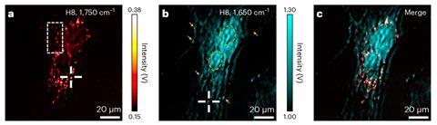
In the new research, Zhang and colleagues started from the label-free technique mid-infrared photothermal (MIP) imaging, which probes the response of objects to pulses of infrared radiation by studying the way they diffract continuous-wave visible light – a material’s refractive index changes when it’s heated. MIP usually has a resolution of around 300nm – around 10 times greater than traditional IR imaging simply because of visible light’s shorter wavelength – but certainly not super-resolution.
The researchers noted that, as expected, pumping an object with a pulsed IR laser produced modulations of the frequency of the probe laser with the same interval. These modulations were not simply sinusoidal oscillations, however: there were several high-frequency harmonics of the fundamental frequency. ‘It was something of an accidental discovery,’ says Zhang, ‘because I was thinking that … if we added these different harmonic orders together maybe we could increase the signal-to-noise ratio, but then I saw that the different harmonic orders looked different, so I was thinking “What is going on?”.’ The answer turned out to be that, as the object is not uniform but instead has a detailed internal structure, the pump pulse modulates the refractive index experienced by the probe laser in a complex, spatially-dependent way. Combining the effects of multiple harmonics of the fundamental modulation frequency therefore allows the researchers to perform super-resolution observations of the internal structure of the object.
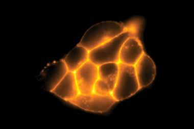
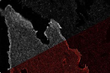
In one demonstration, they examined features only 86nm in size in living yeast cells. The researchers call the technique photothermal relaxation localisation or Pearl microscopy.
‘I think it’s a really cool paper,’ says microscopist Eric Potma of the University of California, Irvine in the US. Photothermal imaging has proved extremely useful for imaging biological samples since its introduction around 10 years ago, he says. ‘This is done in such a simple and elegant way that … you don’t have to change your hardware, your microscope, even in principle the software – you just have to make sure your lock-in amplifier can handle higher harmonics. With the flip of a switch you should be able to identify biologically meaningful features that previously remained hidden with existing implementations of photothermal IR microscopy.’
References
P Fu et al, Nat. Photon., 2023, DOI: 10.1038/s41566-022-01143-3


No comments yet