Spectroscopic techniques allow scientists to look over the shoulders of old masters. Emma Stoye reports
The scientific scrutiny of art dates back hundreds of years. Accounts from as early as the 16th century describe collectors grinding up and analysing the creations of painters and sculptors – sometimes for restoration, but often just out of interest. Most of the techniques now used have been borrowed and adapted from other fields – x-rays, for example, were used on paintings shortly after they were developed for medical imaging.
Nowadays, the main challenge is to investigate without causing damage. There is unfortunately no silver bullet for this, and different pieces require different techniques because of the vast range of materials used to make paints (see The colourful science). There may be limitations, but art historians are increasingly recognising the value that science can bring, says Tim Smith from Renishaw, a company whose spectrometers have been used to study and authenticate works of art.
‘Having a technique that provides chemical information without subjectivity or ambiguity is of huge benefit to museums and galleries,’ he explains. ‘It can help to determine the pigments, their source, their date, and identify whether any changes have been made.’
Detecting fraud, for example – which was once completely reliant on experts scrutinising the style of a painting – has become easier. Smith recalls that one of Renishaw’s Raman spectrometers was recently used to expose a fake Chagall painting that made international news. ‘The blue pigment was identified as phthalocyanine blue and not the expected Prussian blue,’ he says. ‘The watercolour is said to date from 1909–10, but phthalocyanine blue wasn’t available as a commercial dye until after this date.’
X-ray vision
But probing pigments is about more than just sorting the real from the fake. It can tell us a great deal about the way a piece of art was put together, giving historians a glimpse into the artist’s thought processes. ‘When you look round in a museum, you’re only seeing the final surface – nearly all artists have re-used their canvas to sketch and test different ideas,’ says Joris Dik of Delft University in the Netherlands. ‘Just a few microns below the surface there is a wealth of information on how that painting was created. If you can visualise these substructures, you can almost get an idea of what the artist was thinking as he was painting it, layer by layer.’
Dik, who trained first as an art historian then as a chemist, has worked with paintings from great masters at some of the world’s most famous galleries. Like his predecessors, he uses x-rays to detect heavy metal pigments below the surface of paintings, but this approach has been adapted over the years. Classic x-ray tomography (used to detect broken bones) can only detect very heavy metal pigments like white lead oxide, which tends to obscure parts of the painting.
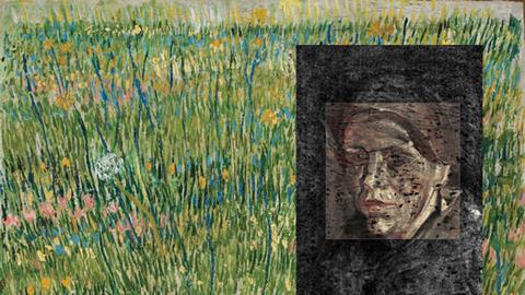
X-ray fluorescence (XRF) spectroscopy, on the other hand, involves firing a beam of high-energy x-rays at the painting, which are absorbed and then re-emitted at characteristic wavelengths by different elements in the paint. ‘Using x-ray fluorescence, we can penetrate the surface and look at the layers underneath and tell their elemental composition – and through that also get an idea of the colour,’ says Dik. He was part of a team that used XRF to map the presence of mercury (found in the red pigment vermilion), zinc (zinc white) and tin (Naples yellow) in van Gogh’s painting Patch of grass, revealing a lost portrait of a woman’s face that the artist had painted over.1 The same technique was also used to reveal Rembrandt’s indecisiveness when painting Syndics of the drapers’ guild, when buried layers of white lead showed he had moved one of the figures to four different locations before finally deciding and painting over the rest.
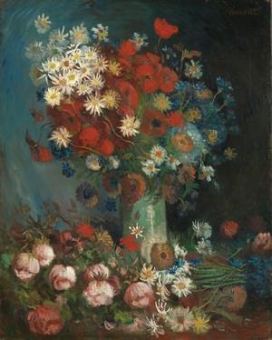
In one of his most memorable discoveries, Dik recalls that XRF analysis led to a painting being re-authenticated as a van Gogh after sitting in a museum storage room for nearly 40 years.2Still life with meadow flowers and roses had been described as ‘artistically dubious’ by critics. There were serious doubts over its authenticity, as the canvas was larger than most of van Gogh’s flower still lifes from the 1880s and the foreground was uncharacteristically ‘busy’ with an extra bunch of flowers in front of the vase. It was dismissed as ‘artist: anonymous’ in 2003.
When Dik and his team came to revisit the painting they discovered it had been painted on top of another, depicting two men wrestling. This matches up to some of van Gogh’s correspondence from his time at the art academy in Antwerp, Belgium, in 1886, when he wrote to his brother that he had painted ‘two nude torsos – two wrestlers’. No other wrestler painting exists, and the pigments – zinc white and mercury-based vermillion – are materials van Gogh was using at the time. It appears he simply recycled the canvas, adding some extra flowers in the foreground to cover up one of the figures.

‘The visualisation of the scene of two wrestlers was instrumental in re-attributing the painting to van Gogh because he had written about it,’ says Dik. ‘Seeing that is like standing behind the artist and looking over his shoulder.’ The painting was re-authenticated in 2012 and put on display alongside van Gogh’s flower pieces in the Kröller-Müller museum in the Netherlands.
Looking deeper with lasers
Though they can tell us a lot about the pigments present in a painting, Dik admits there are limitations to the techniques he uses. ‘One of the things that we’re not good at is getting depth sensitivity,’ he says. ‘We see differences in lower layers because of chemical differences, but there’s not one good technique that gives us an idea about the stratigraphy.’ This is problematic when there are lots of layers underneath a painting, or when the same paints are used on different layers.
‘Right now, to understand these things, even with extremely expensive paintings, conservators take a scalpel to it,’ says chemist Warren Warren from Duke University in the US. Tiny chips taken from non-prominent parts of the painting or the margins of existing cracks can be examined in cross section under a microscope to look at the layers.
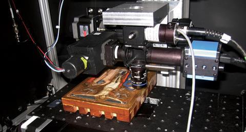
But Warren and his group have found a non-destructive way to image paintings in three dimensions, by adapting a laser-based technique they originally developed for medical imaging. Femtosecond pump–probe microscopy uses carefully timed laser pulses to excite the molecules in a sample to a higher energy state. As the molecules fall back down again, they emit signals that, as with XRF, can be used as a ‘fingerprint’ to identify them. But the technique can be used on organic as well as metal-based pigments and – crucially – the lasers can be focussed at precise depths, allowing pigments to be mapped in 3D.
Warren’s group is primarily interested in using pump-probe microscopy to look through layers of skin and detect cancerous tissue. But during a trip to the National Gallery while at a conference in the UK, it occurred to Warren that it might work well on the pigments in paintings as well as those in skin.
‘The melanins and haemoglobins in skin have very broad absorption spectrum. So, the first question we asked is, if we do nothing at all to modify our laser system, what would we be able to look at?’ says Warren. ‘Our system works in the near infrared and so blue pigments are pretty well suitable for its principles.’
Experimenting with different paints, the group found the technique could clearly distinguish between purple colours created by layering red paint on top of blue and those made by mixing the two together. They then teamed up with conservators at the nearby North Carolina Museum of Art and tested the technique out on a 14th century painting of the crucifixion by Puccio Capanna. In a purple section of the painting depicting Mary’s robe they detected a layer of ultramarine, made from ground-up lapis lazuli that at the time was ‘more expensive than gold’, says Warren. On a different purple area – the angel’s robe – they could see layers of ultramarine blue paint and a red glaze, as well as white iron oxide and gold leaf. Their observations were verified by samples.3
‘This shows we can get the same information without taking a scalpel to the painting,’ which provides a damage-free way to study how the painting was created, says Warren. This kind of information can help historians distinguish parts painted by a master from those painted by a student, or show how an old painting has been touched up over the years.
The group are now working on extending the range of pigments and colours their system can detect. ‘By simply changing the wavelengths of the laser we use, we can probe just about any pigment that we’re interested in,’ says Warren. They also want to develop a more portable system which would enable them to visit galleries further afield. At the moment, the art has to come to them.
Into the terahertz world
While analysing paintings on canvas can be difficult, it is nothing compared to the challenge of examining wall paintings, including the murals and frescoes that adorn the walls of churches and palaces around the world.
Like oil paintings, the majority of frescoes have evolved over time – they may have been restored, repainted or even plastered over if the owner has a change of heart. Revealing lost layers is fraught with challenges. The work usually can’t be taken off the wall, which eliminates transmission techniques, which require access to both sides, or large immovable equipment. And methods like XRF may also be unsuitable, because the radiation can’t penetrate through thick paint and plaster.
You can image paintings even through five or six millimetres of plaster
Bianca Jackson
But the expanding field of terahertz imaging offers some hope. This uses pulses of long-wavelength terahertz radiation – which sits between infrared and microwaves on the electromagnetic spectrum – to penetrate through opaque materials and give a depth profile of the different layers. ‘When you have a material with multiple layers, you’re going to get reflection at each of the interfaces between layers,’ explains Bianca Jackson, of the University of Reading in the UK, who uses terahertz radiation to image art and historical artefacts. ‘It is then possible to separate out these layers and create discrete images.’ The technique is comparable to medical ultrasound and CT scans, she adds, which also work by detecting pulses reflected at the boundaries between different materials.
Terahertz imaging has been used in other fields – including airport security scanners – for several years. Jackson first began to explore its potential for investigating art when she was a graduate student at the University of Michigan in the US. She and a colleague tested the technique out in a makeshift art studio, experimenting with different paints and materials. ‘Some pigments offer a much better contrast than others, but terahertz is a hundred to a thousand times longer wavelength than infrared, which means you have better penetration,’ says Jackson. ‘I found you could image paintings even through five or six millimetres of plaster.’
This makes the technique ideal for analysing wall paintings. One of Jackson’s first successes was revealing Neolithic wall art covered by centuries of plaster at an archaeological site in Catalhoyuk, Turkey.4 Another exciting discovery came during a trip to the Louvre gallery in France. While Jackson wasn’t able to image underdrawings of the Mona Lisa, because the red ochre (iron oxide) pigments have a very low contrast, she did discover part of the original Roman-era image underneath a fresco – Trois hommes armes de lances – revealing the extent to which it had been ‘restored’ by its forger–collector Giampietro Campana in the 19th century. Conservators had already suspected there was a picture underneath the surface, says Jackson: ‘XRF had detected pigments that were not on the surface.’ But they had failed to get an image because of the thick paint and plaster. After many hours of painstakingly scanning the fresco, the outline of a face was revealed beneath the left hip of one of the figures in the painting.
This success makes terahertz imaging one of the newest additions to the art investigator’s toolbox, a toolbox that is continuing to expand as equipment becomes more sophisticated, portable and robust. With an almost infinite supply of art all over the world – only a fraction of which has been investigated in detail – there is no doubt plenty left to discover. Now, thanks to a small number of groups who are bridging the divide between science and art, we are starting to see art more clearly than ever before.
Picasso’s paints
Restorers are often keen to find out exactly what materials artists have used, because it can help them look after pieces better, safely repairing damage and keeping them at optimal conditions. Volker Rose and his team at Argonne National Laboratory in Illinois, US, recently used high intensity x-rays to show that Picasso used common house paint, rather than traditional artist’s oil paint, to make the white parts in some of his paintings.5

Historians had suspected this for a long time, as the basic appearance of house paint differs considerably from oil paint. By teaming up with conservators at the nearby Art Institute of Chicago, Rose and his group were able to study the impurities in the white zinc oxide using a high intensity x-ray nanoprobe at the laboratory’s Advanced Photon Source, and compare them to samples of old house paint they bought on eBay. From this they identified the paint Picasso used as belonging to the French brand Ripolin.
Read more in our chemistry and art theme issue
References
1 J Dik et al, Anal. Chem., 2008, 80, 6436 (DOI: 10.1021/ac800965g)
2 Van Gogh: new findings, Wbooks, Zwolle/Van Gogh Museum, Amsterdam, 2012
3 T E Villafana et al, Proc. Natl Acad. Sci.USA, 2014, 111, 1708 (DOI: 10.1073/pnas.1317230111)
4 G C Walker et al, Opt. Express, 2013, 21, 8126 (DOI: 10.1364/OE.21.008126)
5 F Cansadio and V Rose, Appl. Phys. A: Mater. Sci. Process., 2013, 111, 1 (DOI: 10.1007/s00339-012-7534-x)


No comments yet