
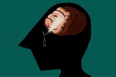
The connections between brain cells have been mapped with unprecedented molecular resolution using a new light-microscopy technique that swells and blows up brain tissue without significantly distorting it. The researchers believe the technique, which most labs could carry out, could provide new insights into neuroscience and could also be useful in other tissues.
The resolution of traditional light microscopy is constrained by the diffraction limit to be no more than half the wavelength of visible light, limiting it to about 200nm at best. This is insufficient to resolve dense cellular structures such as neurons’ synaptic connections in the brain, whose finest details are just tens of nanometres in size. Researchers have therefore turned to electron microscopy. The Machine Intelligence from Cortical Networks (Microns) project recently produced a structural map of the myriad neuronal interconnections in a millimetre of mouse visual cortex this way.
Electron microscopy provides little information on the molecules present in the structures imaged, however. This currently involves correlating structural electron microscopy with fluorescence microscopy. ‘This requires dual-domain expertise and equipment, and there’s a mismatch in the resolution … so the pinpointing of the molecules is not as precise as one might wish,’ says Johann Danzl at the Institute of Science and Technology Austria near Vienna.
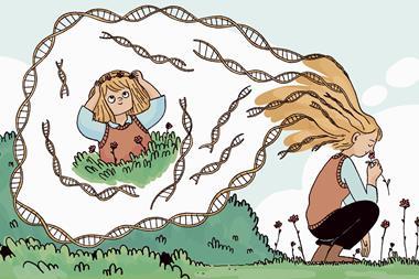
In the new work, Danzl and colleagues achieved the necessary molecular resolution with light microscopy by physically enlarging the samples using swellable hydrogels. This technique, called expansion microscopy, has existed for around 10 years, but it has never previously been possible to expand a tissue sample with sufficient scale and fidelity to reconstruct neuronal connections.
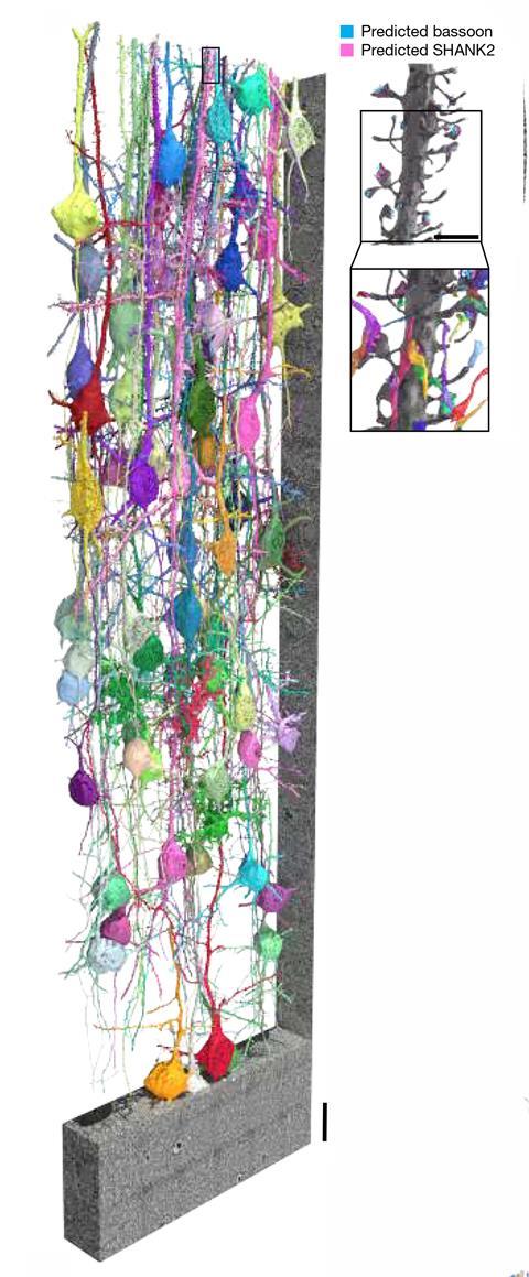
The researchers developed a new protocol in which a solution containing one type of hydrogel monomer (acrylamide) was distributed through the vascular systems of mice together with the chemical fixative. After the animals’ brains were removed and sliced, they were treated with the co-monomer sodium acrylate to form a swellable hydrogel. The researchers then heated the tissue and used other chemicals to disrupt its mechanical cohesiveness before applying multiple further hydrogels to the tissue, increasing its size by a factor of 16. The neurons could then be analysed using light. The researchers could also add antibodies to label specific molecules.
A deeper look
In collaboration with Viren Jain at Google Research, the team used deep learning techniques similar to those previously used by the Microns group to trace and identify individual neurons. They manually traced the paths of neurons through multiple images and used these as training data for the deep learning algorithm. After three rounds of optimisation, the algorithm could make reliable predictions of neurons’ synaptic connections independently.
The molecular labelling data provides information that would be difficult to obtain through electron microscopy, says Danzl. ‘There are certain molecules associated with excitatory synapses and certain molecules associated with inhibitory synapses,’ he explains. ‘Excitatory synapses are pretty straightforward to see purely from the structural channel; inhibitory synapses, on the contrary, are less conspicuous in the structural channel, and there it’s extremely helpful to have the molecular labelling to unambiguously say that this is an inhibitory synapse.’ Various other aspects of synaptic physiology could also be labelled, he says. The researchers have termed the technique light-microscopy-based connectomics (Liconn). As it does not require specialised equipment, the researchers believe it should be widely accessible to neuroscience labs and could be extended to analyse other organs.
‘This is the first paper that illustrates that there’s a way to look at synaptic connections between neurons that is also totally compatible with also measuring the molecules in the sample,’ says neuroscientist Forrest Collman at the Allen Institute in Seattle, part of the Microns collaboration. ‘The difference between a good dataset and a bad dataset is often driven by the histology portion of the protocol, not the imaging portion. This protocol is the first one where, after you take a picture, what you get looks like the structure has been preserved in a dense way that lets you trace the individual axons and dendrites.’
He reveals that his own group has collaborated with Danzl since the Austrian group developed the protocol. ‘Some of our initial attempts to follow the protocol produced samples that weren’t pretty,’ he says. ‘We communicated back and forth and found out what things we were doing wrong and what steps we were not following exactly as they had intended. After a couple of rounds of iteration with them, we were able to reproduce their results and get really good pictures.’
References
MR Tavakoli et al, Nature, 2025, DOI: 10.1038/s41586-025-08985-1


No comments yet