Self-assembling protein tubes can be shaped into a flexible, branching network that can support growing cells
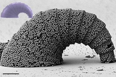
An international team of researchers has made tubular protein-based structures that can be shaped into a network by manually pulling out new branches from existing tubes. The structure is self-healing and bioactive, and can support the growth of human cells for tissue engineering.
Alvaro Mata of Queen Mary University of London and collaborators in Israel, Portugal and Spain combined molecular self-assembly with direct physical manipulation to draw out the tubes from a protein-based membrane. The basic fabric is a composite of a synthetic polypeptide based on elastin – a stretchy protein found in connective tissue – with a small amphiphilic peptide that has water-soluble head and a fatty, insoluble tail. Such peptide amphiphiles (PAs) have been shown previously to be capable of self-assembling in water into structures such as nanofibres and gels, which have themselves been explored as potential scaffolds for cell growth.
When Mata and colleagues added the PA solution to the elastin-like protein (ELP), the PA formed a roughly spherical blob enclosed in a soft membrane, which is composed of an ELP-PA complex. On their own, the elastin molecules collapse into compact blobs in water. But when the PAs bind to them through non-covalent interactions such as hydrogen bonds and electrostatic interactions, they can be opened up in a manner akin to protein denaturation, as the PAs solubilise hydrophobic parts of the polypeptide. The ELP-PA complex may then aggregate to form a membrane. Electron microscopy showed that this has a multilayered structure, like lasagne, with each layer just a few nanometers thick.
If the membrane is touched – by a glass rod, a finger, more or less anything – it changes shape over the course of a few minutes, forming an opening where the touch occurred. If it is touched top and bottom, it opens into a tube that eventually bridges the top and bottom of the solution, still with the PA solution inside. In much the same way, if a glass rod touches the wall of the tube, a new tube forms there which can be gently pulled out like a branch. The extrusion process looks rather like nylon threads being pulled from the interface of the two polymerising liquids.
Any touch will open the membrane, says Mata. ‘If we were to inject the peptide drop into a swimming pool of the ELP, and then inside touch it with say your five fingers, you will open five openings around the blob, each the diameter of your fingertips.’ The researchers have extruded tubes as narrow as 800µm, but Mata says he is confident that they could be made narrower still. And as the tube is extruded, it can quickly self-heal any ruptures in the membrane.
Shapeable scaffolds
Given the biocompatibility of elastin, Mata and colleagues figured their tubular scaffolds might be good hosts for growing living cells. They found that mouse stem cells and human endothelial cells from umbilical tissue will successfully colonise the structures in a few weeks. This means that the ‘shapeable’ tube networks might be used to make complex scaffolds, perhaps even acting as a vascular network, without the need for moulds or templates. They are stable for months under cell culture conditions, and flexible and relatively strong for a self-assembling material – slightly more so once coated with cells.
The tubes ‘may be useful as blood vessels or neuronal conduits to guide nerve cell growth’, says Rein Ulijn of the City University of New York. ‘A major advantage of this system is the possibility to manipulate the tube in real time and with spatiotemporal control, which means that diameter, length, membrane elasticity and even branching may be controlled.’ He notes that the membranes are chemically distinct on the inside and outside, which raises the possibility of giving the two sides different receptiveness to different types of cell, which could be of particular use for growing nerve conduits and blood vessels.
The researchers aren’t yet sure exactly why the membrane-enclosed blob of PA solution can be so readily transformed into a tube. But they suspect it is a dynamical process in which any disruption of the membrane, for example by a physical contact, allows the peptide amphiphiles to diffuse through to the water surface, where they self-assemble into a film that then widens the rupture, rather like a film of soap molecules spreading on the water surface. The whole process is highly sensitive to the nature of the molecular interactions: PAs with only one less lysine residue no longer make the multilayered membrane when combined with ELPs, but instead a denser fibrous aggregate.
Stephen Mann of the University of Bristol, UK, says that, while the idea of generating membrane-bounded polyelectrolyte capsules by droplet immersion in a concentrated solution of the second component is well known, what he finds novel is the dynamical behaviour that allows the tubes to be extended under tension by recruiting more material to the membrane. ‘I would expect the next step to involve an element of rational design using computer modelling and simulations’, he says. This won’t be easy, he cautions: ‘there appears to be a complex interplay of molecular and supramolecular interactions across several length scales.’
References
Inostroza-Brito et al, Nat. Chem., DOI: 10.1038/nchem.2349





No comments yet