Vibrational spectroscopy performed on the nanoscale, opening up investigations into bonding structure
Scientists will now be able to better understand the physical and chemical properties of materials thanks to researchers who have added vibrational spectroscopy to the electron microscope at a spatial resolution of just a few nanometres. The work will enable scientists to detect atoms that were previously invisible to electron microscopy and analyse sensitive materials without damaging them.
Vibrational spectroscopy is a powerful analytical tool for studying the vibrational excitations of molecules inside materials, thus providing insight into their composition and properties. However, such spectroscopy is typically limited to a spatial resolution of 1µm or more.
Now, Ondrej Krivanek, at Arizona State University and president of electron-optical instrument company Nion, together with colleagues at institutions in the US and Canada, has taken vibrational spectroscopy into the nanoscale by introducing it into a transmission electron microscope.
‘The vibration signal has always been present in the electron microscope, but in the typical electron energy spectrum it was obscured,’ says Krivanek. ‘What is surprising to other people in our field is that we’ve managed to improve the energy resolution and other aspects of the technique so much that the vibrational signal is now clearly visible.’
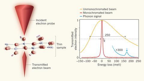
The team reached a spatial resolution of 5nm by developing a new electron monochromator – a device that transmits only a select band of electromagnetic wavelengths – to improve the energy resolution of electron energy loss spectroscopy (EELS) carried out in the electron microscope.
The new technique works by firing a very high energy and narrow beam of electrons – as small as an atom or even smaller – through a finely sliced sample. This makes the atomic nuclei in the sample vibrate and by analysing the minute energy transfers from the electrons to the sample it is possible to observe how the nuclei are vibrating.
‘Being able to do vibrational analysis in the electron microscope with nanometre-level spatial resolution will allow all kinds of new experiments, such as reliably detecting hydrogen, an element that has up to now been essentially “invisible” in the electron microscope, as well as analysing the bonding structure of the hydrogen atoms,’ Krivanek says. Furthermore, the technique has the potential to carry out analysis of radiation-sensitive materials, such as polymers or frozen hydrated biological tissues without damaging them.
‘The experiment itself is a tour de force and opens new fields for electron microscopy,’ comments David Muller, a materials and electron microscopy expert at Cornell University, US. ‘The electron microscope’s existing ability to image atom-sized features inside a material or device has made it invaluable for materials science, but adding vibrational spectroscopy provides information that is more familiar to chemists – basically imagine Raman spectroscopy from an atomic-sized probe.’
‘This is a brand new capability and there is much work ahead. In particular, we will be working on improving both the energy and the spatial resolution,’ says Krivanek. ‘Our ultimate goal is to be able to detect and analyse the vibrations of a single atom.’


No comments yet