New technique could improve tumour diagnosis and treatment
A new automated fluorine-18 labelling procedure could make this useful radioisotope easier to use in medical imaging.1
Positron emission tomography (PET) is a nuclear medicine technique that provides a non-invasive way to diagnose and design treatment for tumours. The technique monitors how positron-emitting radionuclides bound to biological targeting molecules accumulate in the body. The radionuclide accumulates in cancerous tissue, and its concentration provides both qualitative and quantitative information about the tumour.
Radiochemists often consider fluorine-18 the ideal PET radioisotope thanks to its excellent radioactive decay characteristics – high positron emission, low positron energy and half-life compatible with off-site transport. In the past, incorporating 18F into organic molecules involved high temperatures and organic solvents, making it unsuitable for labelling biomolecules such as peptides and small proteins. However, direct radiolabelling of biomolecules with 18F has recently been developed, and researchers in the US have reported a radiolabelling kit containing all of the reagents in one vial.2
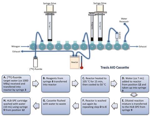
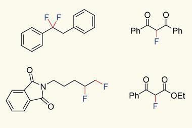
At the Institute of Cancer Research in the UK, Graham Smith and co-workers have now taken this isotope one step closer to the clinic by automating the process. Using two different radiolabelling platforms, they’ve successfully labelled three substrates and achieved over 98% radiochemical purity. This could allow them to produce large batches of radiopharmaceuticals for satellite PET centres around the country.
Automated procedures are a requirement of good manufacturing process-compliant radiopharmaceuticals; enabling batch reporting and record keeping, control and standardisation of processes, and reducing the risk of contamination. ‘Removing the human interaction from a process provides a reproducible production time and a standardised approach to radiosynthesis,’ explains Smith, who believes his technique will allow more widespread use of fluorine-18 in the clinic. ‘There will be a direct patient benefit due to the superior decay characteristics of fluorine-18 over comparable radioisotopes such as gallium-68,’ he adds.
Ralf Schirrmacher, an oncologic imaging expert at the University of Alberta, Canada, is full of praise for the research: ‘The current study impressively attests to the impact and synthetic power of this new radiochemistry, and is a perfect example that out-of-the-box thinking results in innovation, automation and application.’
The team hopes this process will provide a foundation for automating radiopharmaceutical production, enabling huge advances in patient diagnosis and treatment.
References
1. L Allott et al, React. Chem. Eng., 2017, DOI: 10.1039/c6re00204h (This article is free to access.)
2. W J McBride et al, Bioconjugate Chem., 2012, 23, 538 (DOI: 10.1021/bc200608e)


No comments yet