
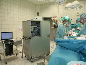
A combination of Raman spectroscopy and sound allows doctors to identify brain tumours while their visual attention remains focused on the surgery.
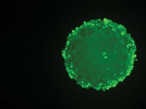
Every cell has a unique Raman spectrum that is like a fingerprint; tumour and healthy brain cells, for example, have different spectra. Raman spectroscopy is ideal to obtain real-time samples because it is non-destructive, simple to operate and does not require sample preparation.
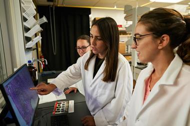
Scientists in the UK have developed a technique that examines brain cells using Raman spectroscopy, and then transforms the spectroscopic data into sounds. Because different types of cancer cells give distinct Raman spectra, a music synthesiser can produce different timbres for each of them. During surgery, doctors can concentrate on the scalpel, while listening to whether they are removing a tumour or healthy tissue.
The team, led Matthew Baker at the University of Strathclyde in Glasgow, tested their invention with a group of volunteers. Participants were able to identify the different timbres and accurately classify (77.1%) up to three kinds of brain cells: healthy cells, metastatic tumour cells and aggressive glioblastoma cells. Baker’s method could bring Raman spectroscopy closer to clinical applications.
References
This article is free to access until 14 December 2016
R Stables et al, Analyst, 2016, DOI: 10.1039/c6an01583b


No comments yet