
Two coumarin compounds with nearly identical structures but contrary fluorescent properties have been isolated from the orange climber plant. One may find use in biomedical imaging due to its unconventional behaviour when aggregated in solutions.
Many traditional fluorescent molecules do not emit light when in solid forms. But aggregation-induced emission (AIE) luminogens show the opposite phenomenon, glowing brightly even in an aggregate state. While many synthetic versions of these have been developed and found use in theranostics and luminescent sensors, they tend to be toxic to the environment, have poor biocompatibility, and are difficult to make.
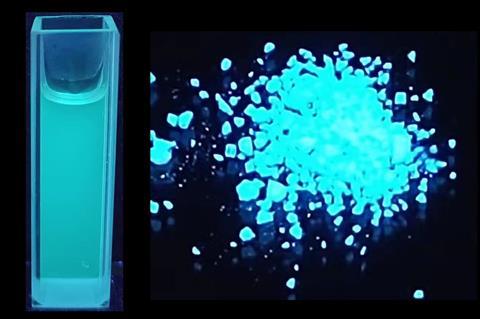
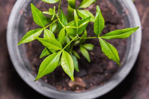
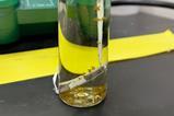
Inspired by the blue fluorescence displayed by the roots of the orange climber plant (also known as Toddalia asiatica or Zanthoxylum asiaticum), researchers from China isolated two isomeric compounds that are the first natural coumarins discovered to be AIE-active.
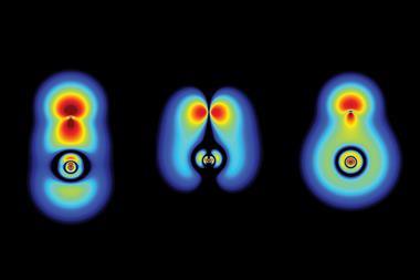
The scientists found that one of the coumarins, 6-methoxyseselin, displays AIE through the typical mechanism of restriction of intramolecular motion caused by its rigid structure and the presence of multiple hydrogen bonds between molecules. But this mechanism does not explain AIE in the other isomer.
Further analysis revealed that 5-methoxyseselin glows brightly in dilute solutions or crystal states but shows a quenching effect in protic solvents like water. The researchers believe this unexpected phenomenon is due to an unconventional electron energy transfer between the solvent and aggregates.
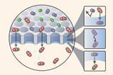
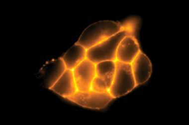
Both coumarins were tested for live-cell imaging applications. While each displayed good biocompatibility and low cytotoxicity, due to its unique water-sensitive fluorescence, 5-methoxyseselin provided wash-free mitochondria staining with minimal background signal. The researchers theorise that the compound disaggregates inside cells and interacts with cellular proteins to switch fluorescence back on, providing a fast and powerful tool for visualising mitochondria in live cells.
References
S-S Chen et al, ACS Cent. Sci., 2023, DOI: 10.1021/acscentsci.3c00012


No comments yet