
Brain organoids – self-organising clusters of human neurons just millimetres across – are bridging the long-standing gap between animal models and human disease, offering new insight into conditions that have resisted decades of research and opening routes to therapies that were previously out of reach
- Brain organoids, or ‘mini-brains’, are 3D structures grown from human stem cells that self-organise to resemble aspects of the developing human brain, offering a more realistic model than rodents or cells grown in flat cultures for studying neurological and psychiatric diseases.
- Researchers can create patient-specific organoids from skin or blood cells, allowing them to investigate disorders such as schizophrenia, Rett syndrome and rare epilepsies, revealing disease mechanisms and neural network abnormalities that are difficult or impossible to study in animals.
- The technology is also being applied to neurodegenerative diseases including Alzheimer’s, with increasingly sophisticated organoids incorporating immune cells, blood-vessel components and other support cells to better reproduce disease processes and test potential treatments such as lecanemab.
- Despite challenges such as the lack of a true blood supply, advances including vascularised organoids, multi-region ‘assembloids’, microfluidic systems and even transplantation into animals are making mini-brains more realistic, while early clinical trials based on organoid research suggest the field is beginning to translate into therapies.
This summary was generated by AI and checked by a human editor
For decades rodent research remained one of the few ways to investigate complicated brain conditions such as Alzheimer’s disease. Yet a mouse brain, slightly smaller than a garden pea, weighs less than half a gram. Mice patently do not have a brain layout like humans.
There were other methods: Neuroscientists could manipulate human neurons in a dish, for instance. But a layer of cells is some distance from the complex network of the 1.4kg adult human brain. Then, brain organoids – or mini-brains as they are sometimes called – arrived.
What are brain organoids?
Just a few millimetres of spherical blob, these tissues have surprised scientists in their ability to self-organise and mirror some features of the foetal brain. ’What we had not anticipated when we first started was how faithful this process is to what happens in the developing brain,’ says Benneth Novitch, a molecular biologist at the University of California, Los Angeles in the US, who creates brain organoids. Clinical advances are now within reach. Mini-brains recently catalysed a breakthrough for a rare brain disease.
An advantage of organoids is that they place cells into a context that resembles a developing brain. They sit in a 3D matrix and better replicate communication networks between the cells. They hold special promise for neuropsychiatric conditions that are impossible to replicate in rodents. ‘Mice don’t get bipolar disorder or schizophrenia. We induce it but it’s not the same,’ says Annie Kathuria, bioengineer at Johns Hopkins School of Medicine in Baltimore, US.
More than 90% of drugs entering clinical trials for neurological and psychiatric conditions fail to receive approval, highlighting the need for better disease models. Costly failures at late stages have triggered sporadic industry pullbacks. Meanwhile, organoids have become more sophisticated, with improved cellular diversity and longer culture times. They can complement research in animals.
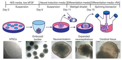
The starter ingredient for mini brains is the conversion of skin or blood cells into human induced pluripotent stem cells (iPSC), which can develop into any cell type. A special brew of growth factors and inhibitors will shunt them towards an early version of the central nervous system. A seminal advance in 2013 grew cerebral organoids in a gel protein mixture and was led by Juergen Knoblich at the Institute of Molecular Biotechnology in Vienna, Austria. The development was unguided, with a somewhat messy but interesting mix of cell types. But researchers also began trying to generate specific brain localities.
Chemical recipes for midbrain organoids were devised, for instance. This brain area is critical in movement, coordination and reward pathways. Dopamine neurons here progressively die in Parkinson’s disease, and the protocol could allow for drug screening or generate donor cells for Parkinson’s therapy. Some firms are already using midbrain organoids to screen new molecules against Parkinson’s. Other researchers have devised culture conditions to recreate different parts of the early human brain, including a mix of cortical and hippocampal tissues. ‘We’ve learnt a lot about key growth factor signals and we can administer these to developing organoids, but it takes a lot of painstaking work to get conditions just right,’ says Novitch.
Modelling schizophrenia with patient-derived organoids
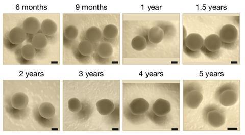
Growing organoids for longer and guiding them with chemical signals let different types of neurons populate the mini-brain, such as excitatory or inhibitory neurons or neurons with specific messengers such as acetylcholine or dopamine. Recently, a group developed human cortical organoids for five years in culture, eventually developing genetic features of human brains after birth.
The early tissue that organoids tend to replicate may offer upsides, because early neuroinflammation is a suspect in adult psychiatric conditions. There is evidence to suggest that infections in pregnant women can generate an immune response that hits certain susceptible brain cells – GABAergic neurons in the cortex. In a proposed trigger for schizophrenia, inflammation or stress later in adolescence again targets these neurons, which help to synchronise brain signalling. ‘Maternal immune activation is a popular hypothesis for neuropsychiatric disorders – so children develop diseases at a later stage because they were exposed to immune fire in utero,’ says Kathuria.
A major advantage of brain organoids is that ordinary skin cells can be taken from patients and cultured into mini-brains. In a 2020 study, Kathuria and her colleagues turned cells from eight patients with schizophrenia and eight healthy controls and grew them into brain organoids . It revealed molecular and functional abnormalities in organoids from schizophrenic patient cells and postmortem brains from patients.
In California, Novitch and his colleagues generated organoids derived from patients with Rett syndrome, a rare genetic condition that impairs movement and cognition and may include epilepsy. They fused excitatory and inhibitory neurons and then probed the patient-derived mini brains with imaging and electrical signalling analyses. The organoids from Rett patients developed epileptic-like activity in the neural network – important because epilepsy is a network abnormality. ‘We’re now trying to create brain-like structures that show changes in neural circuit activities in epilepsy and look at how the cells in an organoid are communicating with one another,’ says Novitch.
Recently, the Novitch group developed organoids with a mutation in a sodium channel associated with a rare childhood epilepsy. Children with this condition experience frequent seizures, developmental delays, intellectual disability and autism. In cortical organoids made from patient cells, neurons fired too frequently – mimicking seizures. While in hippocampal organoids, brain rhythms linked to learning and memory were disrupted because of a loss of neurons that policed neural activity. This research would be difficult in animals. ‘Some populations of brain cells are enlarged in humans, but not rodents,’ says Novitch.
He is part of a consortium investigating 250 genes linked to neurodevelopmental and psychiatric disorders using Crispr–Cas9 gene editing in human cell lines, zebrafish and mouse. ‘Rather than relying on patient genetics, we will take neurotypical patient cell lines and put mutations into hundreds of genes linked to these disorders,’ Novitch explains. ‘We will then ask the question – when you differentiate these cells into 2D or 3D culture, what does it change about their development?’
Brain organoids and Alzheimer’s disease: bridging the gap between mice and humans
Neurodegenerative conditions such as Alzheimer’s are big targets for brain research. There are challenges in trying to put this brain condition into a small rodent. The genetics in so-called Alzheimer’s mouse models is also problematic. Less than one in 20 cases of Alzheimer’s are caused by an inherited mutation, often causing early-onset disease. The same proteins are mutated in mice because it triggers brain abnormalities – abnormal plaques of insoluble protein – that look like end-stage Alzheimer’s disease. But the mice themselves do not have the complex sporadic form that make up 95% of Alzheimer’s cases – which simmers for decades before symptoms appear. This might be why many treatments may clear brain plaques in mice but fail to improve patients’ lives. ‘If we get results from animal models [for Alzheimer’s], sometimes we need to validate it using human tissues to make sure it is real,’ says Ranjie Xu, a neuroscientist at Purdue University in Indiana, US, who grows mini-brains to investigate Alzheimer’s.
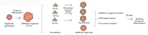
Human brain organoids can help bridge this gap, especially if certain crucial support cells are present.
Inflammation is a hallmark of many brain disorders, including Alzheimer’s, so neuroscientists have worked to include microglia, key immune cells. They respond to injury and influence development. ‘During brain development, there are many unnecessary synapses and they need to be removed. Microglia play a role there,’ says Xu. They also clear cell debris, keep neurons healthy and may stoke inflammation in Alzheimer’s.
Microglia migrate into the brain, early in development, so will not spontaneously develop from neural stem cells. One 2022 investigation showed that microglia boost neurons in midbrain organoids. When Xu put microglia into his organoids, he noted that the microglia seemed to release neurotrophic factors – which can act like brain fertiliser – and the organoids looked healthier. Xu and his colleagues recently developed complex organoids with multiple cell types, including microglia, blood vessels and astrocytes – star-shaped support cells that wrap around blood vessels and neurons. When brain extracts from sporadic Alzheimer’s patients were added to the organoids, plaques and protein aggregates appeared within four weeks, along with impaired neural network activity mimicking the disease state. Treatment with the antibody-targeting treatment lecanemab then reduced the plaque burden.
Nevertheless, there is a paradox in using prenatal brain tissue to investigate ageing conditions like Alzheimer’s and Parkinson’s. ‘Most people will admit that age is the biggest risk factor for any of these diseases,’ says Rusty Gage, geneticist and neuroscientist at the Salk Institute in California, US. Even cells taken from elderly patients will revert back to younger neurons in brain organoids. Gage worked on this ‘conundrum’ by taking patient skin cells and trying to keep their biological age. His group generated induced neurons straight from skin cells using transcription factors and small molecules, instead of first generating stem cells and then neural progenitors. ‘I’m interested in ageing per se and the risk that ageing brings to the brain,’ says Gage.
He subsequently made induced neuronal spheroids, which are not conventional organoids but clumps of neuronal cells. ‘Organoids are dynamically maturing cells. Spheroids are converted from fibroblasts and hold their age,’ Gage explains. Those spheroids generated from Alzheimer’s patients showed neuronal degeneration, plaque accumulation and metabolic dysfunction.
Solving the supply problem
Mini-brains – often the size of blueberries – do not have blood to deliver nutrients and oxygen. The result is dead zones when organoids expand beyond a few millimetres and cells suffer metabolic stress. ‘Neuronal organoids would be happier and healthier if they had an endothelial support system,’ says Kathuria. Blood vessel cells do not originate with neuronal tissue in the embryo, so must be introduced. This is important because the cells that line blood vessels – vascular endothelial – become wrapped with supporting cells called pericytes and astrocytes to create the blood brain barrier. Research has revealed that endothelial cells release lactate when feeding on glucose, which is needed by pericytes, spotlighting how all these cells work as a team.
At the University of Galway in Ireland, biomaterial chemist Mihai Lomora and his colleagues packaged endothelial cells in hydrogel droplets made of an extracellular matrix that degrades over time. This encapsulated growing brain organoid from day eight, with the mini-brains fed endothelial growth factors and a commercial soup of extracellular matrix. ‘The vascular cells want to join up and form a network and they begin to degrade this biomaterial,’ says Siobhan Crilly, a neuroscientist in Galway.
Some organoids even produced their own sack of spinal fluid
By day 40, the organoids contained endothelial cells and neurons, as well as signs of pericytes, astrocytes and microglia. ‘The vascular network perfused the entire tissue,’ says Crilly. She has an interest in using such organoids to study haemorrhagic stroke, which she says is vastly understudied and underfunded because the models are poor, as well as reducing the use of lab animals. A comparison of organoids revealed that those with a vascular mesh were healthier and had less cell death in the centre. There was an up to threefold reduction in apoptosis – cell suicide – compared to non-vascularised organoids.
Others have worked on a supportive milieu for cells. Neurologist Eva Feldman at the University of Michigan in the US joined with bioengineers to generate a matrix made from human fibronectin supported by a porous polymer scaffold to better mimic the natural conditions of a developing brain. Organoids showed greater cell diversity, improved maturation of glial support cells and more robust long-term growth. During a Covid-19 lockdown, some of the organoids were left to grow longer than anticipated, with surprising results. ‘Some organoids even produced their own sack of spinal fluid,’ recalls Feldman. ’This was very similar to human cerebrospinal fluid, which is quite amazing.’
Another strategy is printed 3D scaffolds to replicate diffusion in vascular networks. The tubular channels placed into human midbrain organoids succeeded in warding off hypoxia and dead zones and had more physiologically relevant features, including network activity and neuron maturing. The engineered organoids delivered more regular pharmaceutical responses, such as changes to neuron firings after fentanyl exposure. Meanwhile, Kathuria has patented her recipe for a biological extracellular matrix from human and animal material which she calls the ‘secret sauce’. The result, she says, is organoids that show more mature neurons and faster electrical firings, as well as stepped up expression of the right genes in hindbrain blobs. Kathuria and her colleagues went on to create a multi-region organoid with mid- and hindbrain organoids brought together on day 20 with endothelial cells that integrated with neural tissue and supported development.
Brain assembloids model psychiatric disorders
The result of cultivating brain regions in a dish and bringing them together is often called brain assembloids. A landmark study in 2017 by Sergiu Pasca at Stanford University in California, US, brought together dorsal and ventral forebrains, followed by a combination of cerebral cortex, hindbrain and skeletal muscle tissue in 2020. More recently, Pasca and his colleagues put together a four-part assembloid from stem cells that included somatosensory, spinal, thalamic and cortical organoids. They used this to investigate pathways involved in sensory disorders and proposed that it could be useful to screen therapeutics for pain.
All brain organoids presently lack blood coursing through their tissue, but there are groups working with microfluidics and miniature pumps to try to replicate the rhythmic pluse that starts with a heartbeat. ‘These chip-like devices are advancing but there’s not a lot of publications,’ says Gage. ‘Lots of clever engineers are trying to push blood through tissue and companies are involved because they want to use these for drug screening.’ Others foresee more complex organoids with blood vessels but also other body tissues.
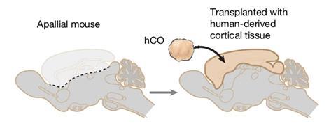
There is another way to get a human organoid perfused with nutrient-rich blood – place a human mini-brain into a living animal. This was first reported in 2018 when Gage and his colleagues put human mini-brains into the brains of adult mice, with neuronal connections and blood vessels forming. A subsequent study by Pasca and colleagues melded human brain organoids into the brains of newborn rats, which then received sensory inputs from the rat brain and even triggered sensory responses in the human cells. The evidence suggested that the human neurons were part of the rat brain, and organoids obtained from patients with Timothy syndrome (a rare genetic disorder) revealed abnormalities in the rat brain that were tougher to detect in culture.
Therapies based on brain organoids are now edging closer to patients. Two trials are coming from the lab of Alysson Muotri at the University of California, San Diego in the US, which sent patient brain organoids to space. ‘The most exciting use of organoids is when we age them at the International Space Station,’ says Muotri. ‘While this also happens in 2D cells and animals, the combination of 3D anatomy and accelerated ageing is really unique.’ One trial is an experimental gene therapy for a rare neurodevelopmental disorder (Pitt-Hopkins syndrome) that saw its first patient treated in February 2026. The second is an intervention for Rett syndrome with an existing HIV drug (lamivudine) – a patient trial began in December 2025.
Human brain organoids are ‘crossing a translational threshold,’ revealing disease mechanisms inaccessible in animals and driving therapeutic discovery, Muotri recently wrote. ‘The field is moving towards more closely resembling the complexity of the human brain in terms of cell types,’ adds Feldman. Organoids can bridge the perilous gap between more traditional lab animals and early tests in patients. ‘It’s not going to get up off the desk and walk away, but it is mimicking really crucial aspects of the human brain that we haven’t been able to look at,’ says Crilly.
Anthony King is a science writer based in Dublin, Ireland










No comments yet